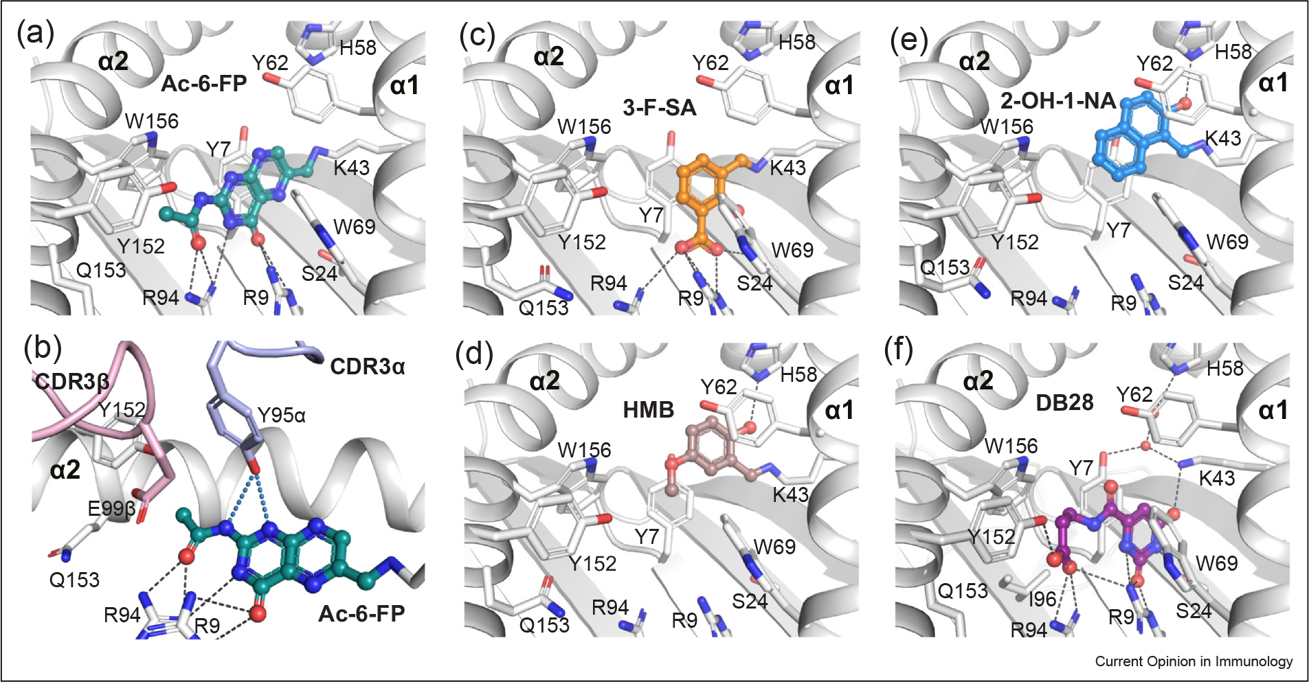
Figure 4.

MR1 presentation of small-molecule MAIT nonstimulating ligands. (a–b) Capture of Ac-6-FP ligand (deep-teal sticks) within the MR1-binding cleft (a) and its interaction with the CDR loops of MAIT TCR (b). (c–f) Docking of diverse chemical identities aside from vitamin-B derivatives within the MR1 groove: (c) 3-F-SA (orange), (d) HMB (brown), (e) 2-OH-1-NA (marine), and (f) DB28 (purple). Coloring as in Figure 3 2. Crystal structures of MAIT–MR1 ligands: Ac-6-FP (PDB: 4PJ5), 3-F-SA (PDB: 5U6Q), HMB (PDB: 5U2V), 2-OH-1-NA (PDB: 5U16), DB28 (PDB: 6PVC), and NV18.1 (PDB: 6PVD), used in figure.
