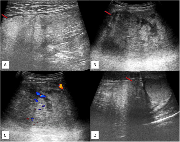
Figure 1. USG images (A-D) of the inguinoscrotal region depict a solid mass with heterogeneous echotexture in the right inguinal region. The mass extends into the right scrotal sac, reaching up to the upper pole of the right testes, and shows internal vascularity.
The USG image reveals a solid mass with heterogeneous echotexture in the right inguinal region, where the superior margins could not be well assessed (red arrow) (A), extending into the right scrotal sac (red arrow) (B), displaying mild internal vascularity (C), and reaching up to the upper pole of the right testis (red arrow) (D).
USG: ultrasonography

