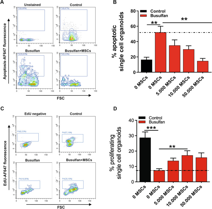
Fig. 8.
MSCs promote regeneration of busulfan-induced damage in intestinal epithelium by regulating the proliferation and apoptosis pathways. A Apoptotic cell death analysis by flow cytometry, of busulfan-treated single cell small intestine organoids stained with Alexa Fluor (AF) 647-conjugated annexin V after co-culture with or without MSCs. B Effects of co-culturing busulfan-treated organoids with 0, 5000, 10,000, and 50,000 MSCs on the percentage of apoptotic single cell organoids are shown. C Proliferation analysis by flow cytometry, of busulfan-treated single cell small intestine organoids stained with EdU antibody after co-culture with or without MSCs. D Effects of co-culturing busulfan-treated organoids with 0, 5000, 10,000, and 50,000 MSCs on the percentage of proliferating single cell organoids are shown. Results are shown as means ± SEM of organoid donor 1 and organoid donor 2 co-cultured with at least 3 MSC donors. ** p < 0.01 and *** p < 0.001 as compared to control (Kruskal–Wallis test)

