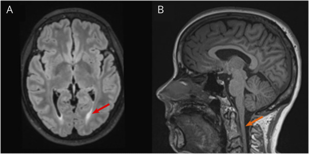
Figure 2. Proband MRI Images.

(A) Transverse brain MRI indicating posterior periventricular white matter changes (red arrow). (B) Sagittal MRI showing diffuse atrophy of the cervical spinal cord (orange arrow).

(A) Transverse brain MRI indicating posterior periventricular white matter changes (red arrow). (B) Sagittal MRI showing diffuse atrophy of the cervical spinal cord (orange arrow).