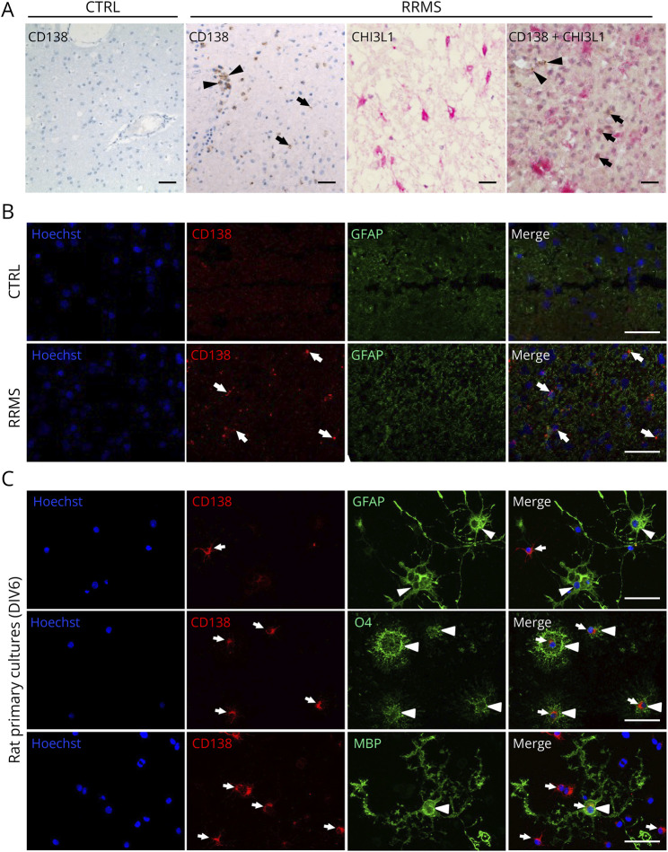
Figure 5. Expression of CD138 in Control and RRMS Human Brain and Rat Primary Cultures.
(A) Immunohistochemistry of brain tissue showing predominant expression of CD138 in cells located in the perivascular spaces (arrowheads) and a sparse expression in brain parenchyma (arrows) of RRMS brain but not in the CTRL brain. CHI3L1 is strongly expressed in astrocytes (pink) from RRMS brain but shows no colocalization with CD138 (brown). Scale bar: 100 µm. (B) Immunofluorescence labeling showing a high expression of GFAP in activated astrocytes of the RRMS brain when compared with the CTRL brain. There is no colocalization of CD138 (arrows) and GFAP in the RRMS brain. Scale bar: 100 µm. (C) Immunostaining of CD138 (arrows) in rat primary cultures of OPCs at 6 DIV showing a stronger expression of CD138 in immature (O4+) than in mature (MBP+) oligodendrocytes and its absence in astrocytes (GFAP+) (arrowheads). Scale bar: 100 µm.

