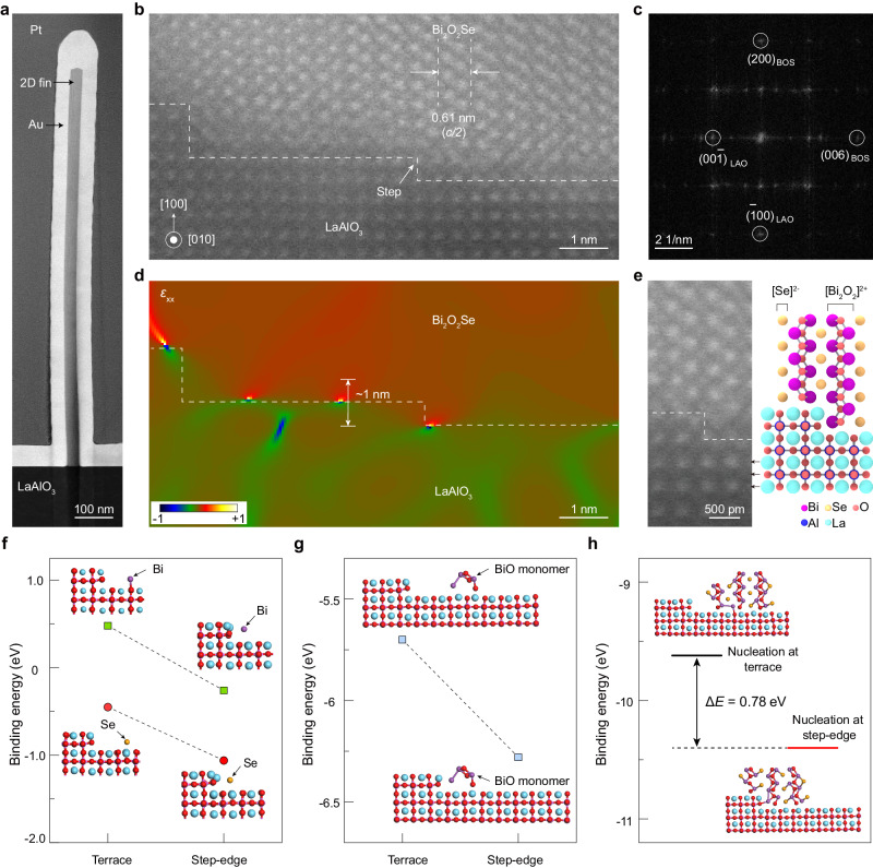
Fig. 2. Structure characterization and nucleation mechanism of vertical 2D fin grown by ledge-guided epitaxy.
a Cross-sectional low-magnification transmission electron microscopy (TEM) image of a vertical Bi2O2Se fin grown by ledge-guided epitaxy. b Cross-sectional high-angle annular dark-field scanning transmission electron microscopy (HAADF-STEM) image showing clear steps on the surface of the substrate, where the fin nucleated. The dashed line means Bi2O2Se/LaAlO3 interface. c Fast Fourier Transform (FFT) diffraction spots of (b). d Strain mapping (ɛxx) estimated from a filtered version of the panel (b). e High-magnification HAADF-STEM image with atomic resolution and corresponding schematic of Bi2O2Se/LaAlO3 interface. The dashed line represents actual interface. f, g The binding energies and optimized structures of Bi/Se atoms (f) and Bi-O monomers (g) adsorbed at the step edge and the terrace of the LaAlO3 substrate, respectively. h The binding energies and optimized structures of a 2D Bi2O2Se nucleus at the step edge and terrace, demonstrate that nucleation at the step edge is energetically favorable. ΔE is the energy difference between two nucleation sites.

