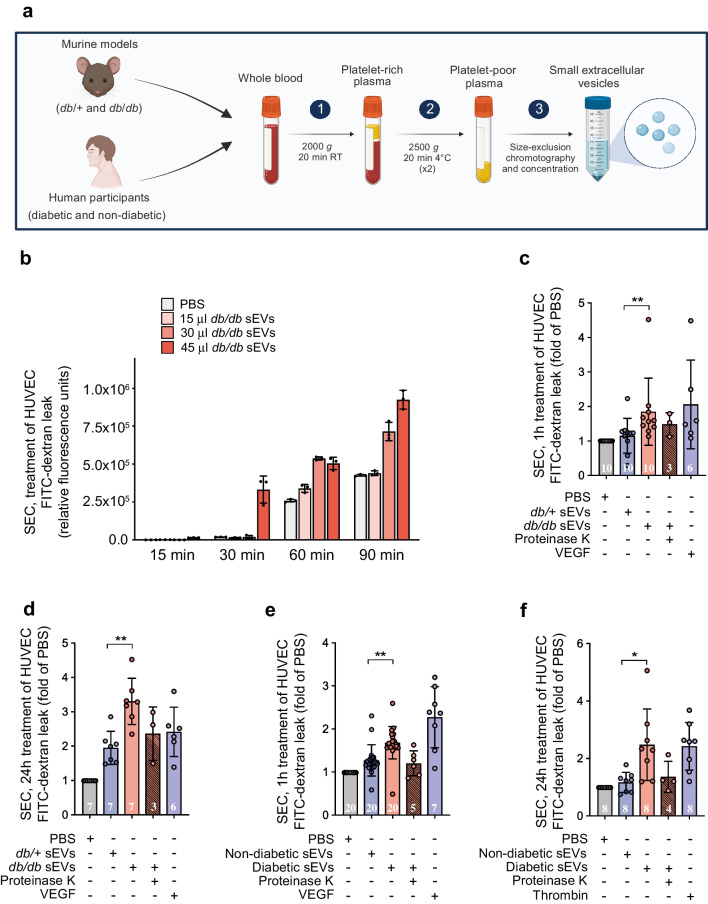
Fig. 3.
sEVs isolated by SEC from diabetic mouse or human plasma induce rapid and sustained increases in endothelial permeability. (a) Schematic detailing the SEC protocol for the enrichment of sEVs from murine and human plasma (see ESM Figs 2, 3 for further details and quality control metrics) (created with BioRender.com). (b) Kinetics of transwell permeability in HUVEC confluent monolayers treated with various amounts of SEC-isolated db/db plasma sEVs for the specified periods, demonstrating that leakage occurs rapidly following treatment in a concentration-dependent manner (n=3 biological replicates). FITC–dextran levels are indicated as relative fluorescence units. (c) Permeability in HUVECs following 1 h treatment (acute) with SEC-isolated sEVs (db/+ or db/db mice) from an equal volume of plasma. (d) Permeability in HUVECs following 24 h treatment (chronic) with SEC-isolated sEVs (db/+ or db/db mice) from an equal volume of plasma. (e) Permeability in HUVECs following 1 h treatment (acute) with SEC-isolated sEVs (diabetic or non-diabetic humans) from an equal volume of plasma. (f) Permeability in HUVECs following 24 h treatment (chronic) with SEC-isolated sEVs (diabetic or non-diabetic humans) from an equal volume of plasma. VEGF was used as a positive control in panels (c–e), and thrombin was used as a positive control in panel (f). In (c–f), the number of biological replicates is indicated at the bottom of each bar; data are relative to the PBS control and are displayed as fold of PBS. The data were analysed using ANOVA with Tukey’s post hoc test *p<0.05, **p<0.01. Values are presented as the mean ± SD. RT, room temperature

