Fig. 7.
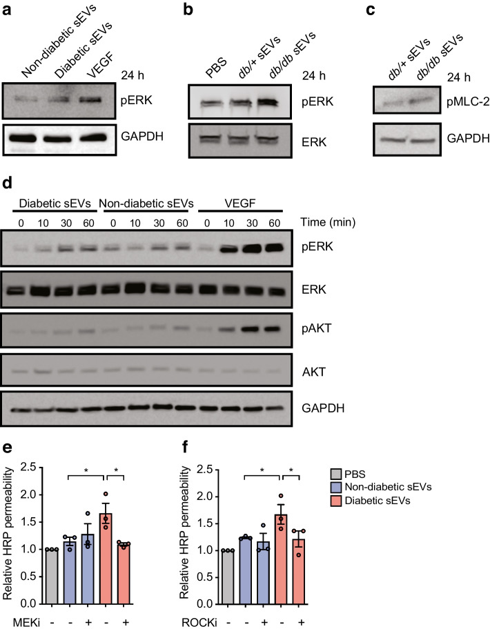
sEVs induce MEK and ROCK signalling, and inhibition of these pathways prevents leakage. (a) Immunoblotting for pERK with GADPH included as a loading control in HUVECs treated with sEVs isolated from an equal volume of plasma from non-diabetic or diabetic individuals. VEGF was included as a positive control for pERK activation. (b) Immunoblotting for pERK and total ERK in HUVECs treated with PBS−/− or with sEVs from db/+ or db/db mouse plasma. (c) Immunoblotting for phosphorylated MLC-2 (pMLC-2) with GAPDH as a loading control in HUVECs treated with sEVs from an equal volume db/+ or db/db mouse plasma. (d) Time course analysis of ERK and AKT activation by immunoblot in HUVECs treated with sEVs from an equal volume of plasma from diabetic or non-diabetic individuals, or with VEGF as a positive control. pERK levels were increased after 30 min of treatment with plasma sEVs from diabetic individuals, with the increase being greater than that resulting from treatment with plasma sEVs from non-diabetic individuals. (e, f) HUVECs were pre-treated with a MEK inhibitor (SL327) (e) or a ROCK inhibitor (Y-27632) (f) and permeability was assessed in response to treatment with sEVs from non-diabetic or diabetic individuals (SEC-isolated from an equal volume of plasma, 1 h, HRP). While MEK or ROCK inhibition did not affect leakage in cells exposed to sEVs from non-diabetic individuals, these inhibitors blocked the increased permeability in response to sEVs from diabetic individuals (n=3 biological replicates). Data in (e, f) are presented as the mean ± SD. ANOVA analysis was performed; *p<0.05. Representative experiments are shown for all immunoblots. MEKi, MEK inhibitor; ROCKi, ROCK inhibitor

