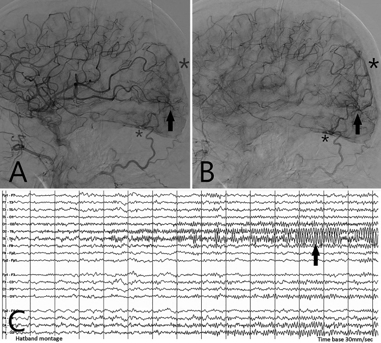
FIG. 1.
Images illustrating the correlation among cerebral hyperperfusion, early venous drainage, and focal seizure activity. A patient with hemianopsia underwent a right common carotid artery DSA (lateral views). During the midarterial phase (A), engorged parietal middle cerebral arteries can be appreciated. Note early capillary hyperperfusion centered on the occipital lobe (arrow). A hint of early venous drainage is already apparent (faint asterisks). In the late arterial phase (B), the early venous drainage is clearly visible (solid asterisks). The main draining veins include occipital surface veins superiorly (large asterisks) and tentorial veins inferiorly (small asterisks). Corresponding surface EEG (C) shows maximum synchronized waves in leads T6 (right posterior temporal) and O2 (right occipital), consistent with a focal occipital lobe seizure. After seizure control with multiple antiepileptic agents, the patient regained his vision, and his cerebral blood flow returned to normal, confirmed by repeat catheter angiography.

