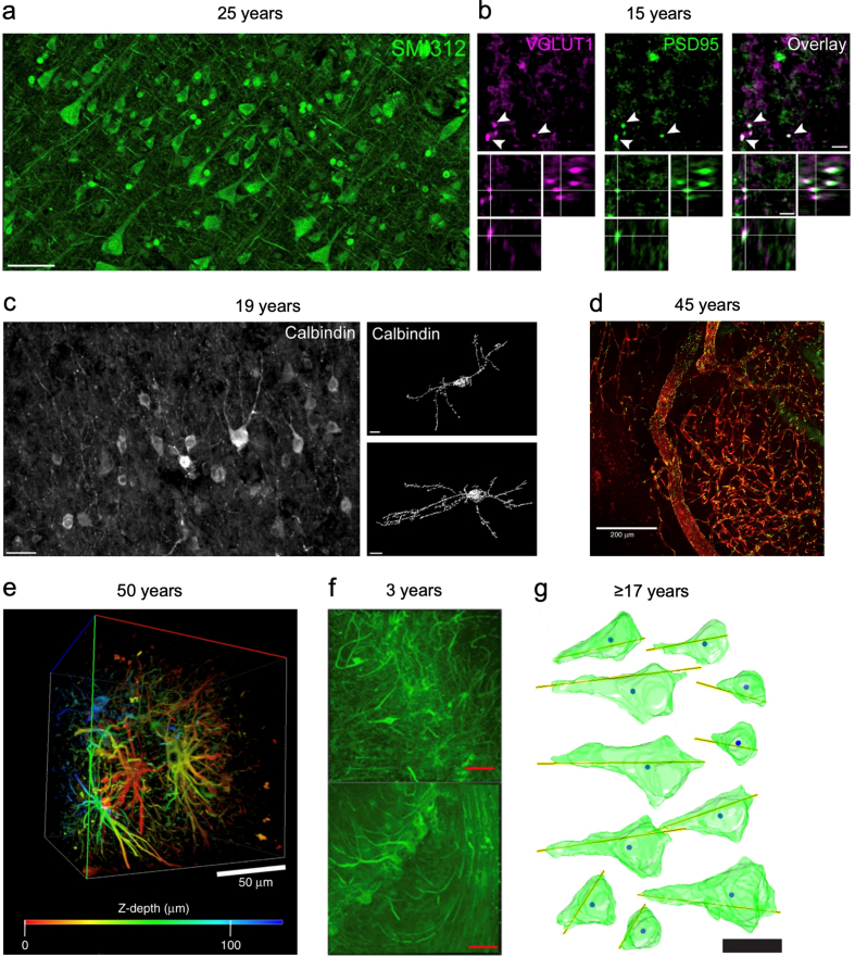
Figure 3. Example images showing morphology preservation from tissue stored long-term in solutions containing formaldehyde.
a-c: Images from (Bouvier et al., 2016). a: Staining for axonal neurofilaments with the antibody SMI312 in cortical sections stored in fixative for 25 years. b: Staining for vGlut1-positive presynaptic boutons (magenta) and PSD95-positive postsynaptic structures (green) and high-resolution imaging allows synapse visualization in cortical tissue stored in fixative for 15 years. c: Staining for calbindin in layers I/II of the cortical tissue stored in fixative for 19 years and 3D reconstruction shows the morphology of calbindin-expressing interneurons. d-e: Images from (Lai et al., 2018). d: Staining for ZO-1 (green) and DyLight 649-labelled lectin (red) allows visualization of blood vessels in cortical tissue stored in fixative for 45 years. e: Color depth-coded, z-stack image of cleared cortical tissue stained with GFAP following 50 years of storage in fixative. f: Image from (Phillips et al., 2016) shows neurofilament H staining in cerebellar tissue from two separate brains (upper and lower) stored in fixative for 3 years each. g: Image from (Larsen et al., 2022) shows a 3D reconstruction of pyramidal cells from tissue stored at least 17 years in formalin, with yellow lines indicating orientation and solid circles the cell centroids. Scale bars = 50 μm (a), 200 μm (b), 5 μm (c), 200 μm (d), 50 μm (e), 100 μm (f), 20 μm (g). All images reproduced under a Creative Commons Attribution 4.0 International License, available here: https://creativecommons.org/licenses/by/4.0/.

