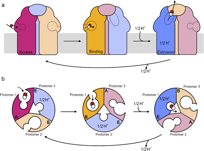
Figure 2.
Cartoon schematic of functional rotation and substrate transport through inner membrane RND transporter proteins. (a) Side-view of the RND protein in the inner membrane. (b) Top-down (from the periplasm) view of the porter domain. For visual clarity, protonation and drug extrusion are considered for a single protomer only. Substrates enter the proximal binding pocket of the Access protomer from the periplasm/periplasmic leaflet of the inner membrane. Substrate binding induces a conformational change in the protomer to the Binding state, and the substrate moves to the distal binding pocket. Protonation then occurs in the relay in the transmembrane domain, inducing a conformational change to the Extrusion state. The periplasmic cleft closes and the exit gate opens, allowing the substrate to exit into the periplasmic adaptor protein (not shown).

