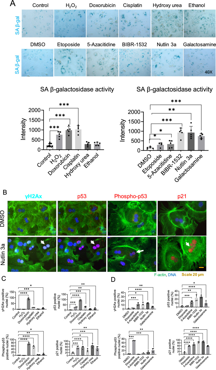
Fig. 1.
Effects of senescence inducers on hepatocytic senescence-associated β-galactosidase activity, p53 and, DNA damage response in primary mouse hepatocytes. A Represented images of SA-β-Gal activity after 24 h of treatment of primary mouse hepatocytes with senescence inducers. The intensity was quantified with Image J. Data were collected from four biological replicates (n = 4) and five technical replicates. The mean of technical replicates was used for statistical analysis. Student’s t-test was used to compare the effect of each treatment with the respective control. Error is shown as standard deviation. (*P < 0.05, **P < 0.005, and ***P < 0.001). B Immunofluorescence images represent senescence-induced DNA damage response markers in primary mouse hepatocytes treated for 24 h in 2D culture, DMSO control panel on top, and Nutlin 3a panel at the bottom. C Drugs soluble in water and, relative quantification. D Drugs soluble in DMSO and, relative quantification. Data were collected from four biological replicates (n = 4) and five technical replicates. The mean of technical replicates was used for statistical analysis. Student’s t-test was used to compare the effect of each treatment with the respective control. The final concentration of 200 μM H2O2, 1 μM doxorubicin, 5 μg/ml cisplatin, 100 μM hydroxy urea, 100 mM ethanol, 10 μM etoposide, 5 μM 5-azacitidine, 20 μM BIBR-1532, 10 μM nutlin 3a, and 1 mM galactosamine were used. Error is shown as standard deviation. (*P < 0.05, **P < 0.005, ***P < 0.001, and ****P < 0.0001)

