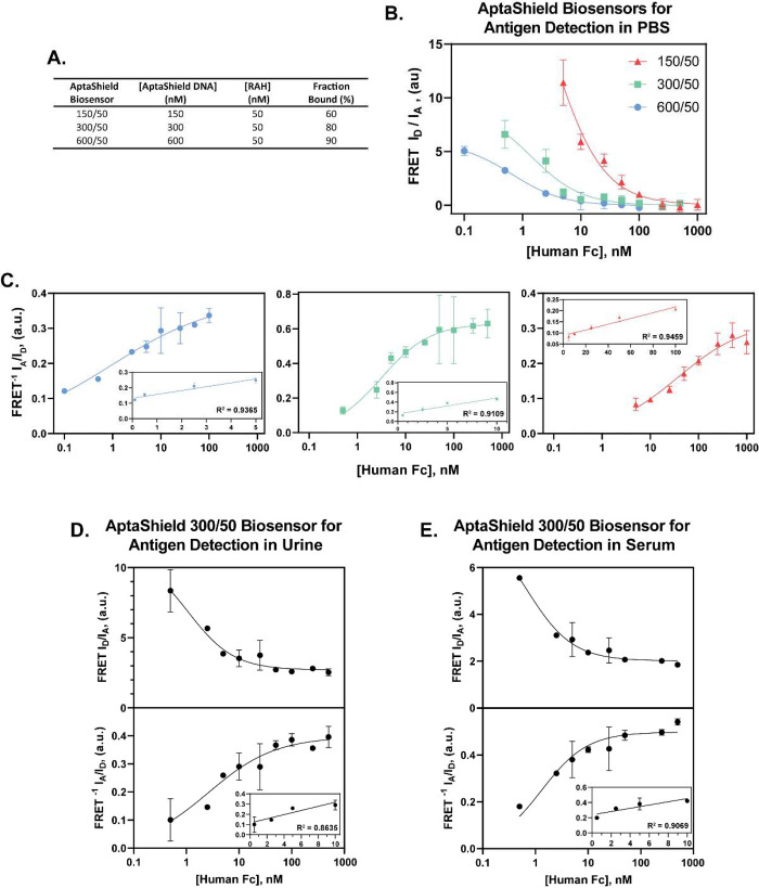
Figure 3.
Analytical performance of the AptaShield biosensor. The proof-of-concept AptaShield biosensor was configured to detect purified human Fc fragment. (a) Different AptaShield biosensors were constructed by varying the ratio between the aptamer and RAH and (b) the baseline subtracted FRET (ID/IA) response curves of these different AptaShield biosensors to different concentrations of human Fc fragment. The nonbaseline corrected binding curves are depicted in Figure S3. (c) Inversion of the FRET response curves (FRET–1, IA/ID) for determination of the Kd Apparent and CLoD of the different AptaShield biosensors. Linear regions of the curves are inset. FRET and FRET–1 response curves of 300/50 AptaShield biosensor to different concentrations of human Fc spiked (d) bovine urine and (e) horse serum. The values are presented as mean ± SEM from 3 to 5 replicas per condition. Statistical analysis was performed using two-tailed t test, and the symbol ** corresponds to p < 0.01.

