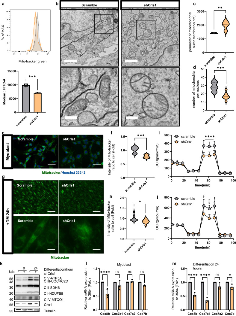
Fig. 5. Crls1 affects mitochondrial structure, leading to the modulation of mitochondrial activity.
a Analysis of mitochondrial mass using flow cytometry. MitoTracker Green staining of myoblasts (gray graph: scramble-infected cell line; yellow: sh-Crls1-infected cell line). Bottom graph: median FITC (green). b Analysis of mitochondrial structure via transmission electron microscopy (left top and bottom panels: infected with scramble; right top and bottom panels: infected with shCrls1; top scale bar: 1 μm; bottom scale bar: 300 nm). c Mitochondrial outer membrane parameter. d Number of mitochondria per nucleus. e Live cell image stained with MitoTracker (left panel: scramble-infected cell line; right panel: shCrls1-infected cell line). Scale bar: 50 μm. f Measurement of the green fluorescence intensity in MitoTracker-stained myoblasts. (n = 7). g Live cell image stained with MitoTracker in the differentiated C2C12 cell line for 24 h in differentiation medium (left panel: scramble-infected cell line; right panel: shCrls1-infected cell line). Top scale bar: 100 μm, bottom scale bar: 12.5 μm. h Green fluorescence intensity of differentiated cells treated with DM for 24 h (n = 11). i Oxygen consumption rate (OCR) in myoblasts (n = 10). j OCRs of differentiated C2C12 cells cultured in differentiation medium for 24 h (n = 4). k Immunoblotting of whole-cell lysates from scramble and shCrls1 myoblasts differentiated for 0 and 24 h. l qPCR analysis of the mitochondrial complex 4 gene from scramble and shCrls1 myoblasts (n = 3). m qPCR analysis of the mitochondrial complex 4 gene from scramble- and shCrls1-differentiated myotubes on Day 1 (n = 3). The graphs present the mean ± SD of n ≥ 3 independent experiments. P values were calculated using an unpaired Student’s t test. *P < 0.05; **P < 0.01; ***P < 0.001; paired t test (a). n.s.: nonsignificant. scramble: untargeted shRNA-lentivirus-infected cell line; shCrls1: Crls1 target shRNA-lentivirus-infected cell line.

