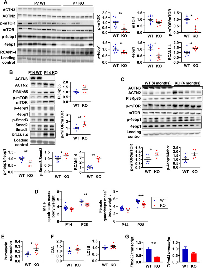
Fig. 1. α-Actinin-3 deficiency alters protein synthesis and breakdown signaling in skeletal muscle from early postnatal development.
(A) At P7, Actn3 KO quadriceps muscles showed significantly reduced activation of mTOR and 4ebp1 but increased expression of RCAN1-4 compared to WT. (B) At P14, Actn3 KO muscles showed increased PI3Kp85, RCAN1-4, and activation of mTOR and Smad3 compared to WT but reduced p-4ebp1/4ebp1 ratio due to increased total 4ebp1 in Actn3 KO muscles. (C) Increased activation of mTOR and 4ebp1 is maintained in adult Actn3 KO muscles relative to WT. (D) At P28, Actn3 KO muscles showed significant reductions in quadriceps mass relative to body weight compared to WT. (E) Puromycin incorporation (indicator of rate of protein synthesis) is increased significantly higher in male Actn3 KO muscles compared to WT. (F) Protein expression of autophagy markers LC3A and LC3B is similar between WT and Actn3 KO muscles; however, (G) transcript expression of E3 ubiquitin ligase genes Fbxo32 and Trim63 is reduced in Actn3 KO. *P < 0.05, **P < 0.01, and ***P < 0.001, Mann Whitney U test. N = 4 to 9 for all experiments.

