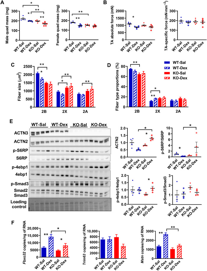
Fig. 5. Female Actn3 KO mice show resistance to muscle wasting induced by prolonged treatment with dexamethasone.
(A) Male WT and Actn3 KO mice show similar levels of muscle atrophy in the quadriceps following 2 weeks of daily dexamethasone administration. In contrast, female Actn3 KO mice showed minimal muscle wasting compared to WT following dexamethasone treatment. (B) Female WT-Dex TA muscles showed significant decrease in maximal force relative to WT-Sal; specific force is similar regardless of genotype and treatment. (C) Female WT-Dex muscles showed reduced fast 2B fiber size compared to WT-Sal and a small increase in 2× fiber proportion (D); there was no change in fiber size or proportions in KO-Dex relative to KO-Sal. (E) KO-Dex muscles show increased activation of protein synthesis markers S6RP and 4ebp1 and a trend for decreased Smad3 activity relative to KO-Sal. (F) Transcriptional changes in Fbxo32, Trim63, and Mstn were assessed by ddPCR in WT and Actn3 KO mice following 7 days of daily dexamethasone injection. WT-Dex muscles showed increased activation of Fbxo32 and Mstn relative to WT-Sal; KO-Dex muscles showed reduced expression of these genes compared to WT-Dex. Scale bar, 50 μm. *P < 0.05 and **P < 0.01 (Mann-Whitney U test). Western blot quantitation values are normalized to the total protein and expressed relative to WT-Sal.

