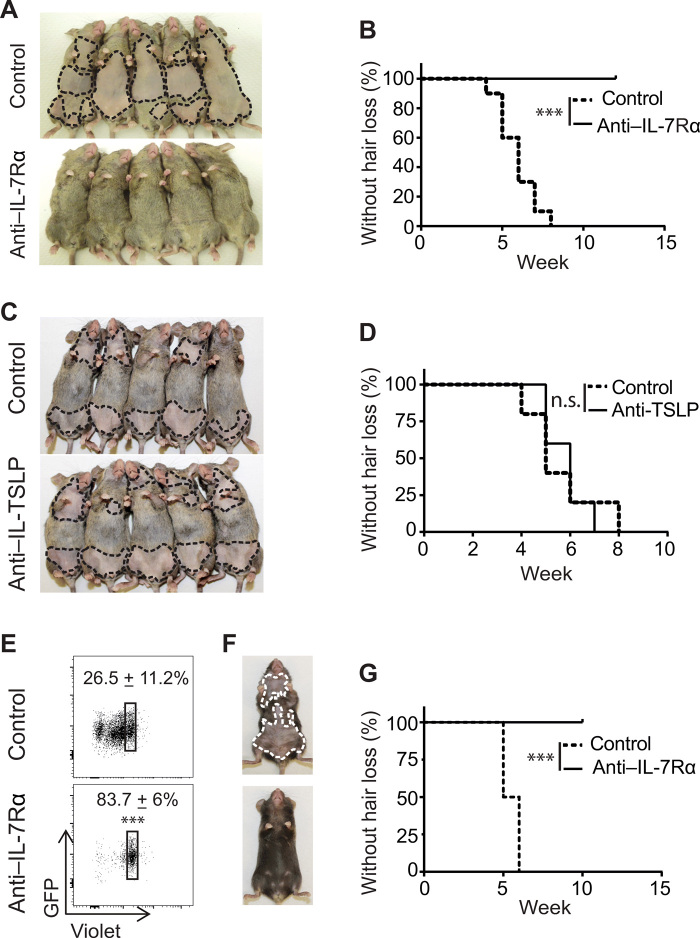
Fig. 3. IL-7Rα blockade prevented development of AA.
(A to D) C3H/HeJ skin-grafted mice were treated with anti–IL-7Rα mAb, anti-TSLP mAb, or control Ab, administrated by intraperitoneal injection two times weekly for 8 weeks, beginning on the day of grafting. Representative images (A) and the incidence of AA onset (B) in skin-grafted C3H/HeJ mice treated with anti–IL-7Rα mAb (n = 15) or control mAb (n = 15) for 8 weeks. ***P < 0.001 (log-rank test). Representative images (C) and the incidence of AA onset (D) in skin-grafted C3H/HeJ mice treated with anti-TSLP mAb (n = 10) or control mAb (n = 10) for 8 weeks. n.s., not significant (log-rank test). (E to G) Purified CD8+ T cells (3 × 10⁶) from 1MOG244 retrogenic TCR B6 RAG1−/− mice were adoptively transferred to B6 RAG1−/− recipients, followed by anti–IL-7Rα or control mAb treatment. (E) The transferred CD8+ T cells were gated on green fluorescent protein (GFP) expression, and the T cell proliferation was measured by CellTrace Violet dye dilution 1 week after treatment. ***P < 0.001 (unpaired Student’s t test). Representative images (F) and the incidence of AA onset (G) in anti–IL-7Rα mAb– or control mAb–treated B6 RAG1−/− recipients (five mice for each group). ***P < 0.001 (log-rank test). Photo credit: Mice pictures taken by Zhenpeng Dai, Columbia University.

