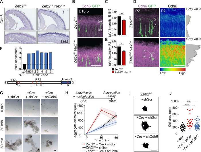
Fig. 8. Cdh6 regulates adhesion downstream of Zeb2.
(A) ISH for Cdh6 in E15.5 Zeb2fl/fl and Zeb2fl/fl NexCre brains. Scale bar, 50 μm. (B to E) Up-regulation and redistribution of Cdh6 protein upon loss of Zeb2. E14.5 Zeb2fl/fl and Zeb2fl/fl NexCre animals were in utero electroporated with GFP and stained for Cdh6 (magenta) at E18.5 (B) and P2 (D). Cdh6 intensity at P2 is shown as a heatmap and gray plot. Scale bar, 50 μm. (C and E) Ratio of Cdh6 in layer I (a) versus CP (b) at E18.5 (C) and P2 (E). N = 3 brains per condition. Unpaired t test. (F) ChIP from E15.5 neocortex shows Zeb2 occupancy at Cdh6 regulatory regions. Red lines mark analyzed sites. Positive control = Ntf3 (fig. S9). (G and H) Cdh6 promotes cell adhesion downstream of Zeb2. (G) Aggregation of E15.5 Zeb2fl/fl and Zeb2fl/fl NexCre neurons transfected with shScr, shCdh6, and Cre as indicated. Scale bar, 100 μm. (H) Average aggregate size. N = 15, 10, and 7 Zeb2fl/fl + shScr; 12, 12, and 15 Zeb2fl/fl NexCre + shScr; and 9, 7, and 4 Zeb2fl/fl NexCre + shCdh6 aggregates at 0, 30, and 60 min. (I and J) Cdh6 regulates adhesion to the extracellular matrix downstream of Zeb2. (I) Attachment of Zeb2fl/fl neurons, transfected with shScr, shCdh6, or Cre as indicated, to laminin-coated surfaces. Adhering cells were visualized after 2 hours by F-actin staining. Scale bar, 15 μm. (J) Lamellipodial spreading. N = 21 Zeb2fl/fl + shScr, 36 Zeb2fl/fl NexCre + shScr, and 41 Zeb2fl/fl NexCre + shCdh6 cells. One-way ANOVA (Kruskal-Wallis test) with Dunn’s multiple comparison test (H and J).

