
Figure 1.
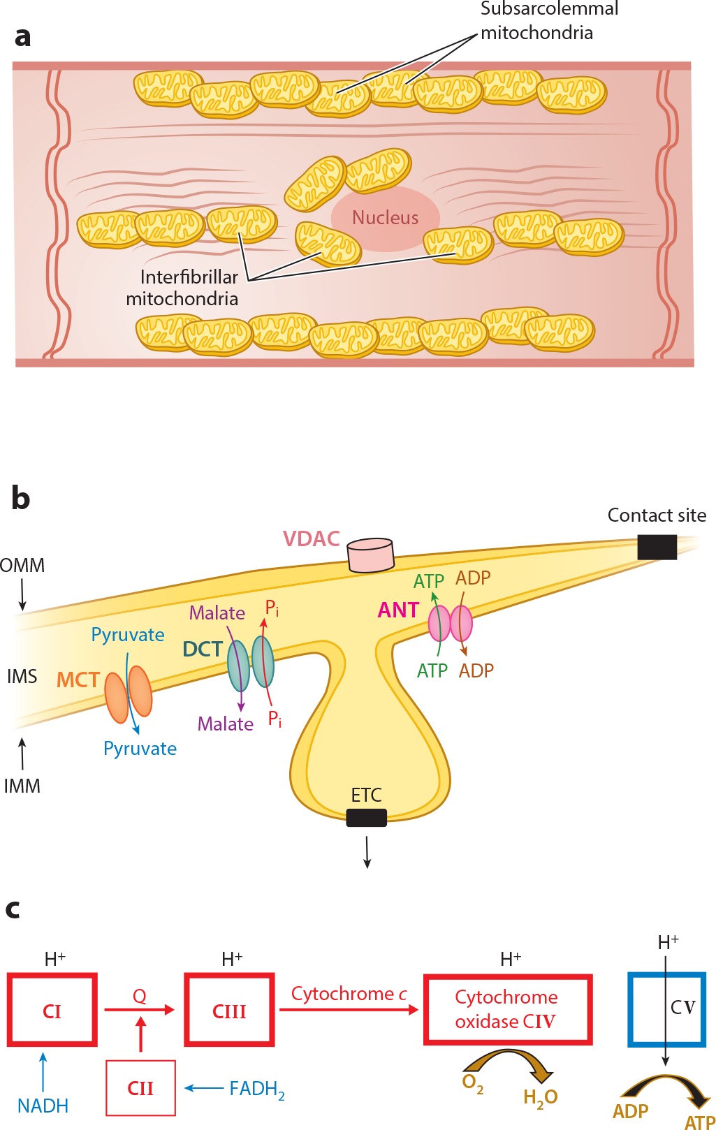
Structure and function of normal mitochondria. (a) The locations of subsarcolemmal and interfibrillar mitochondria in the cardiomyocyte are depicted schematically. The various cellular components are not drawn to scale. (b) Shown are the locations of the voltage-dependent anion channel (VDAC) in the outer mitochondrial membrane (OMM); the electron transport chain (ETC) in the cristae of the inner mitochondrial membrane (IMM) with the adenine nucleotide translocase (ANT), monocarboxylate transporter (MCT), and dicarboxylate translocase (DCT) in the IMM; the intermembrane space (IMS); and contact sites as fusion points of the outer and inner boundary membrane. (c) The ETC. Reduced nicotinamide adenine dinucleotide (NADH) donates an electron to complex I (CI) with flow through to complex IV (CIV) coupled with H+ pumping into the IMS at CI, CIII, and CIV. Reduced flavin adenine dinucleotide (FADH2) feeds electrons into CII with flow to CIV. However, no H+ is pumped into the IMS at complex II. H+ moves into the matrix coupled with the phosphorylation of ADP to ATP by CV. Other abbreviation: Q, coenzyme Q.