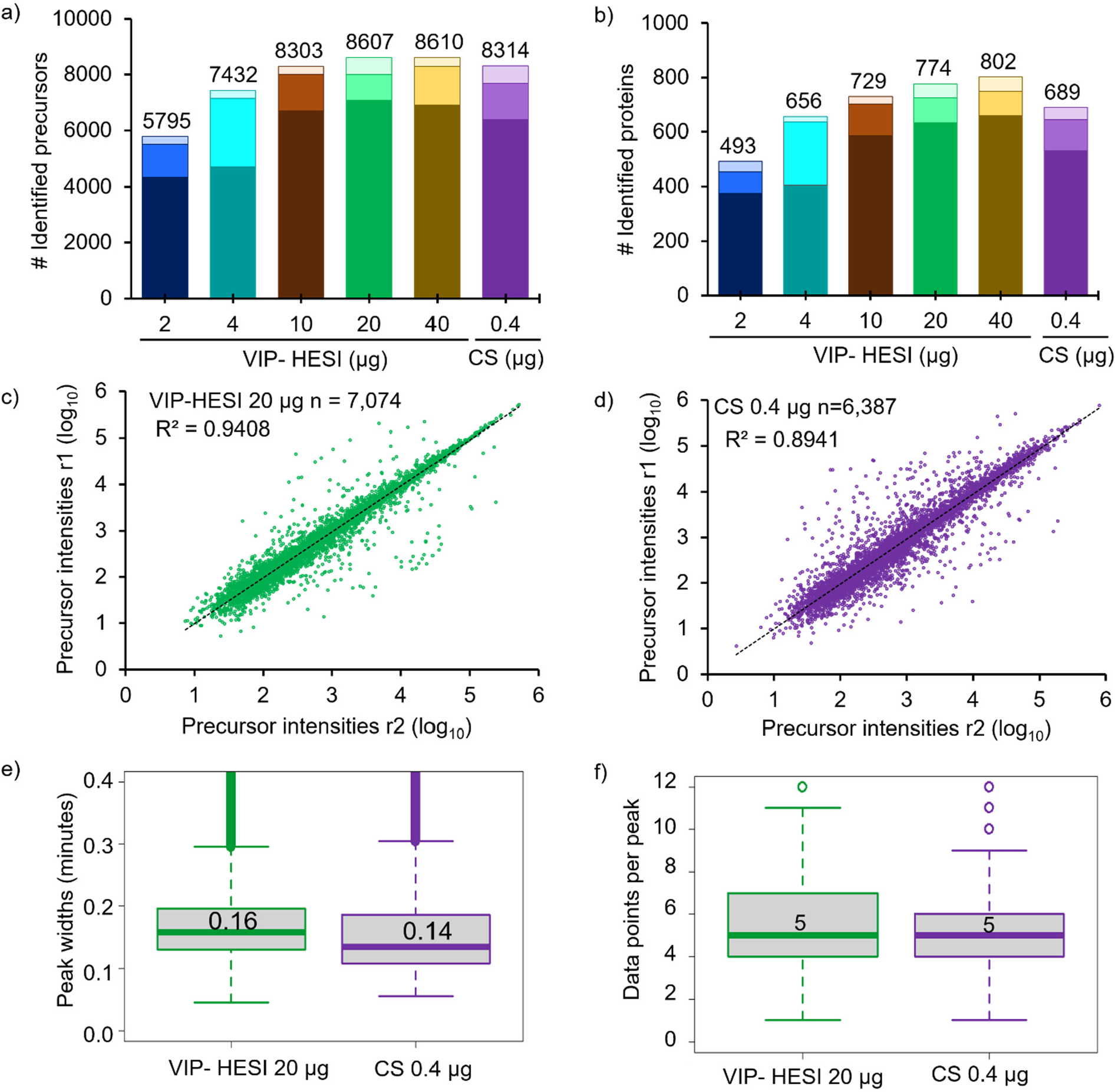
Figure 2.

Comparative performance assessment of VIP-HESI and CS ion source setups using different injection amounts of undepleted mouse plasma samples. a) Number of precursors and b) protein groups identified with different injection amounts of mouse plasma samples analyzed in duplicate with a 45 min 40 μL/min microflow gradient. The darker color bar represents common identification between the replicates whereas the two lighter color bars at the top represents exclusive precursors and proteins identified in each biological replicate at the different sample amounts. Pearson correlation of precursor intensity values obtained from 7,074 and 6,387 precursors that were quantified in both replicates (r1 and r2) using c) VIP-HESI and d) CS source, respectively. e) Distribution of the base peak widths in minutes of precursors identified for VIP-HESI 20 μg and CS 0.4 μg measurements estimated by Spectronaut software. A median value of 0.16 (9.6 s) and 0.14 (8.4 s) peak widths were observed for VIP-HESI 20 μg and CS 0.4 μg injections, respectively. The first and third quartiles are marked by a box with a whisker marking a minimum/maximum value ranging to 0.6 interquartile, and the median is depicted as a solid line. f) Distribution of data points per elution peak for the VIP-HESI 20 μg and CS 0.4 μg measurements estimated by Spectronaut. The first and third quartiles are marked by a box with a whisker marking a minimum/maximum value ranging to 3 interquartile, and the median is depicted as a solid line.
