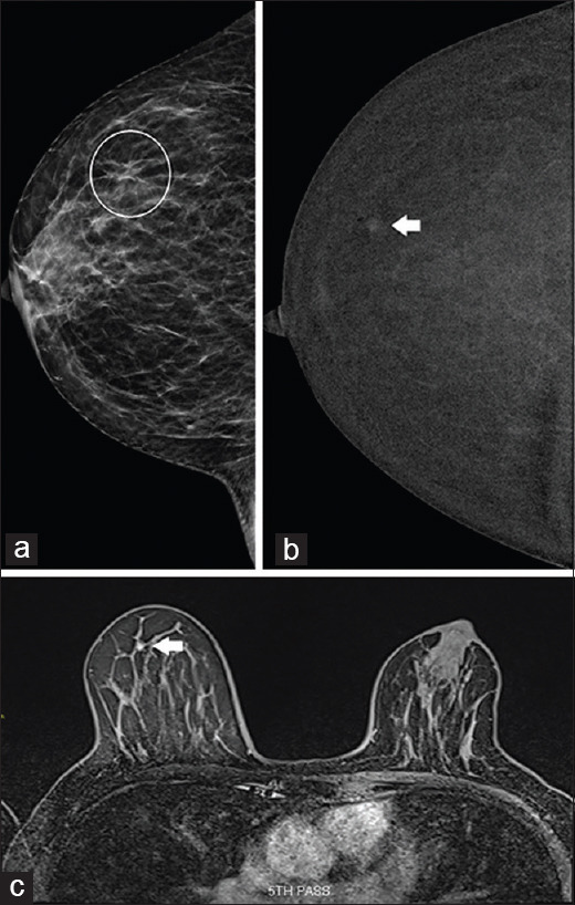
Figure 5.

Case 5: A 59-year-old woman. (a) Full-field digital mammography shows an asymmetry in the outer right breast (circled), visible only on craniocaudal view. (b) Contrast-enhanced spectral mammography (CESM) shows that the lesion was localised to the right upper outer quadrant (arrow). (c) Subsequent MR image shows type I enhancement kinetics, suggestive of a benign lesion (arrow), which confirmed the CESM finding.
