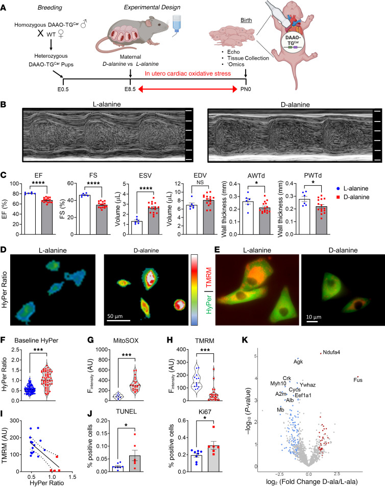
Figure 1. A chemogenetic/transgenic model of neonatal heart failure.
(A) Breeding strategy for heterozygous DAAO-TGCar pups treated in utero. Created with BioRender.com. E, embryonic day; PN, postnatal day. (B) M-mode images of the left ventricle and (C) echocardiographic parameters of ejection fraction (EF), fractional shortening (FS), end-systolic (ESV) and end-diastolic (EDV) volume, and anterior (AWTd) and posterior (PWTd) wall thickness in neonates exposed to d-alanine (0.4 M) (red squares) or l-alanine (0.4 M) (blue circles) from E8.5 to PN0. (D) Baseline HyPer ratio and (E) HyPer and tetramethylrhodamine methyl ester perchlorate (TMRM) images of cardiomyocytes from neonates (PN0) exposed to d-alanine or l-alanine. Quantification of (F) HyPer ratio, (G) mitoSOX, and (H) TMRM fluorescence in arbitrary units (a.U.). (I) Correlation between TMRM and HyPer (r = 0.857, P = 0.0004); dashed lines represent 95% CI. (J) Quantification of TUNEL staining (apoptosis) and Ki67 (proliferation). *P < 0.05, ***P < 0.001, ****P < 0.0001 by unpaired t test. Values shown as mean ± SEM. (K) Volcano plot showing protein abundances in d- versus l-alanine–exposed neonatal hearts; fold-changes and P values are log-transformed. Differentially expressed proteins (P = 0.05; FDR = 0.1) displayed as upregulated (red) and downregulated (blue). Proteins not significantly changed indicated in gray.

