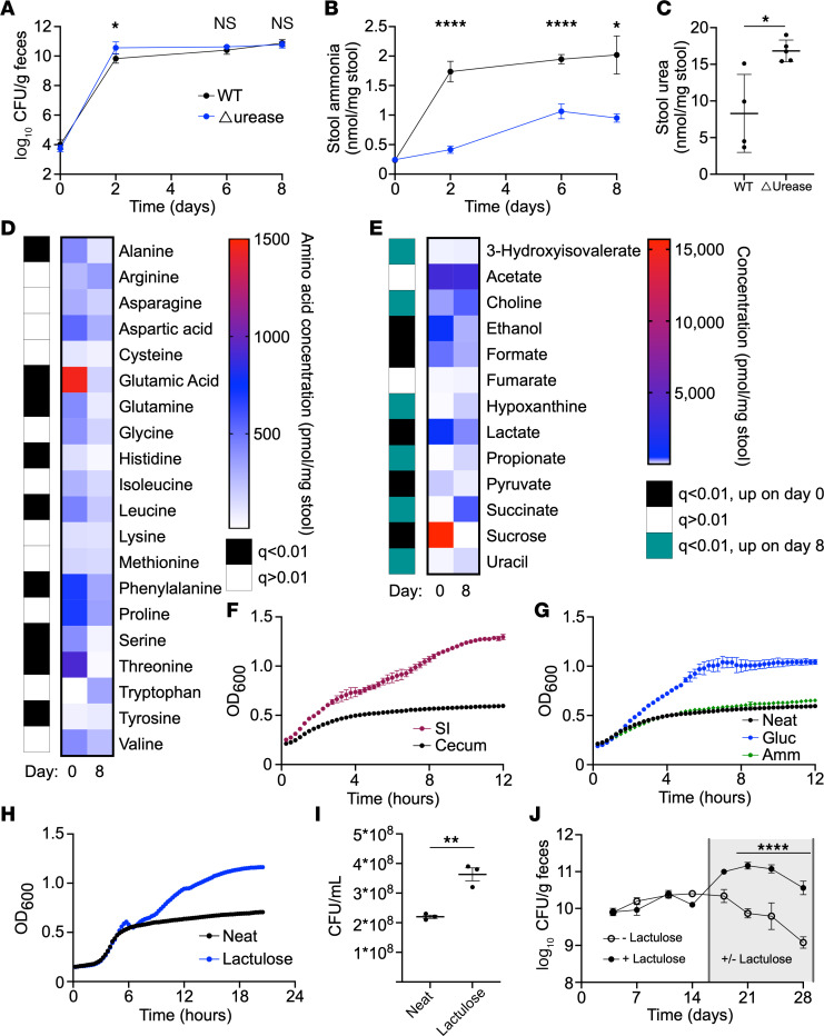
Figure 2. K. pneumoniae colonization is limited by carbon source availability and alters the nitrogen environment in the gut.
(A–E) GF mice were colonized with WT or Δurease K. pneumoniae, and serial stool collections were done throughout the 1-week study. Fecal CFU (A) and stool ammonia (B) were monitored for 1 week after colonization. Fecal urea (C) was tested on day 8. Stool amino acid levels (D) and metabolites (E) were quantified from stool before (day 0) and after (day 8) WT K. pneumoniae colonization. (F) Growth of WT K. pneumoniae in small intestine (SI) or cecal extracts from mice monitored via OD600. (G–I) Growth in cecal extracts supplemented with ammonia (Amm) or glucose (Gluc) (G) or lactulose (H and I) quantified by OD600 and CFU (I). Data for neat cecal extracts are presented in both F and G for reference. (J) Mice colonized with K. pneumoniae were subsequently treated with lactulose in the drinking water or water-only control. Data are presented as the mean ± SEM (A and J) or the mean ± SD (B, C, and F–I). n = 4–5 mice per group (A–E, J) or n = 3 wells per group (F–I). Data represent combined results from 2 independent experiments (A–E) or are from a single experiment representative of 3 independent experiments (F–J). *P < 0.05, **P < 0.01, and ****P < 0.001, by multiple unpaired, 2-tailed t tests with Benjamini Hochberg multiple corrections (D and E), with Bonferroni’s multiple corrections (A, B, and J) or by unpaired, 2-tailed t test (C and I).

