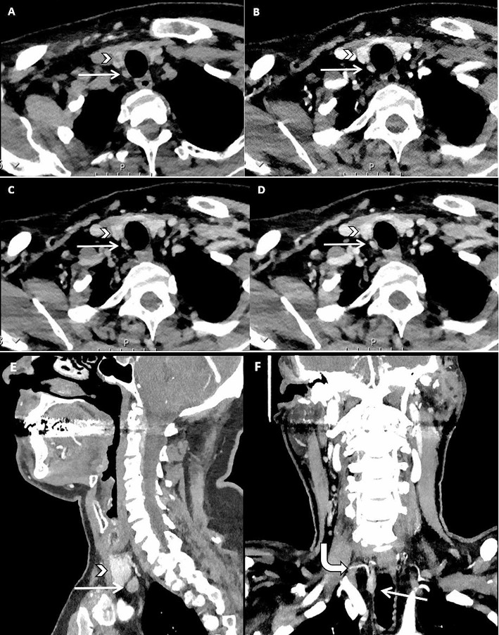
Fig. 3.
62-year-old male with asymptomatic primary hyperparathyroidism. 4DCT showed a hypodense lesion (white straight arrow) dorsally from the right lower quadrant of the thyroid (white arrowhead) on the non-enhanced image (A) with high arterial enhancement (B) and higher washout compared to the thyroid gland on the venous (C) and delayed venous (D) phase. Sagittal reconstruction showed the location of the lesion compared to the thyroid (E). Coronal maximum intensity projection (MIP) showed an enlarged artery (curved white arrow) feeding the lesion, also known as the polar vessel sign (F). The lesion was surgically removed and confirmed by histopathology to be a parathyroid adenoma

