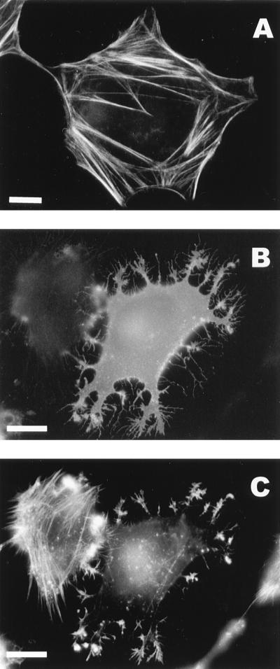
FIG. 3.
Expression of NSP1 causes destruction of actin stress fibers. (A) Oregon-green-conjugated phalloidin staining of stress fibers in a control HeLa cell. (B and C) Lack of stress fibers in a HeLa cell expressing NSP1 3 h after transfection with pTSF1; staining with anti-NSP1 antibodies (B) and Oregon-green phalloidin staining of the same cell (C). Note that stress fibers are present in a neighboring cell, which does not express NSP1. Bars, 10 μm.

