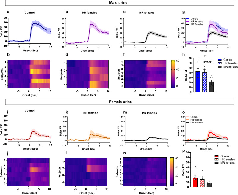
Fig. 7. Activation of the VMHvl nNOS neurons in response to male olfactory cues is specifically disrupted in MR females that were exposed to pubertal stress.
Peri-event time plots and heat maps of the VMHvl nNOS activation in response to male olfactory cues in the control group (a, b), pubertally stressed females that were sexually receptive (HR females) (c, d), and pubertally stressed female that were minimally sexually receptive (MR females) (e, f). h, g Comparison of peak delta F/F nNOS activation in all groups in response to male urine. One way ANOVA (F(2,15) = 6.947, p = 0.0073), followed by Bonferroni’s post hoc test; *p = 0.0116. Peri-event time plots and heat maps of the nNOS neurons responses to female urine in control females (i, j), stressed but sexually receptive females (HR females) (k, l), and stressed females minimally sexually receptive (MR females) (m, n). o, p Comparison of nNOS activation in response to female urine. One-way ANOVA F(2,15) = 2.33, p = 0.131. All subjects were ovariectomized, implanted with an estradiol implant and primed with progesterone 2 to 3 h before the fiber photometry recordings. Control (n = 6), HR females (n = 4), MR females (n = 8). The control group used here is similar to the one used in the previous experiment presented in Fig. 6. All groups were sexually experienced. Data are presented as mean ± SEM. Corrections were made whenever multiple comparisons test is used. Source data are provided as a Source Data file.

