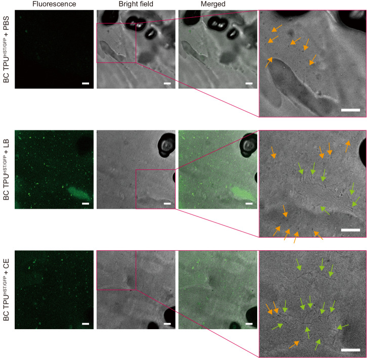
Fig. 6. Genetic engineering of spore-forming bacteria.
Fluorescence, bright-field, and merged images (left to right) of BC TPUHST/GFP incubated in PBS, LB, and CE obtained by CLSM. Rightmost panels are magnified bright-field images to identify the coincidence of rod-shaped vegetative cells with ~5 µm length (green arrows) and particulate spores with ~1 µm length (orange arrows). Scale bars: 10 µm. The experiments were repeated twice, and the representative images were presented. Other images are presented in Supplementary Fig. 16.

