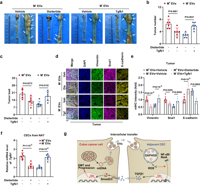
Fig. 8. TGFβ1 expression driven by EV-mtDNA transfer promotes tumor progression in vivo.
a Representative gross view and statistical results of (b) tumor number and (c) tumor load in mice in the disitertide- or Tgfb1-treated murine orthotopic CC model, which received intraperitoneal injections of M+ EVs or M− EVs. n = 6 mice per group. d Representative images of mIHC staining for DAPI (blue), Vimentin (green), Snai1 (magenta), and E-cadherin (yellow) in sections from tumor tissues in the murine orthotopic CC model treated as indicated. Veh Vehicle, Dis Disitertide, Tgf Tgfb1. Scale bar, 100 μm. e The coexpression of E-cadherin, Vimentin, and Snai1 was then evaluated by quantification of the fluorescence intensity. n = 6 mice per group. f The Tgfb1 mRNA levels in CECs isolated from NAT in the murine orthotopic CC model treated as indicated were measured. n = 6 mice per group. g Schematic model showing the mechanism by which the crosstalk between CC cells and normal CECs contributes to tumor progression. Data are means ± SD. One-way ANOVA with Tukey’s multiple comparisons test. Source data are provided as a Source Data file.

