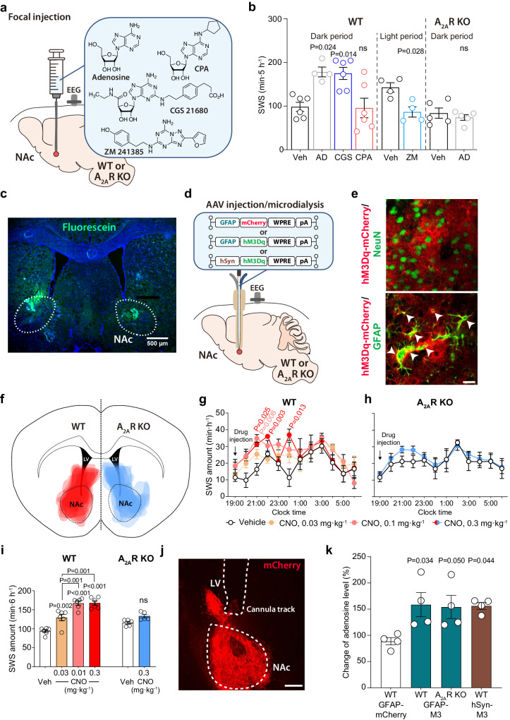
Fig. 1. Activation of NAc A2AR by focal injection of adenosine or stimulation of astrocytes induces SWS.
a Schematic of pharmacologic activation of NAc by focal injection of adenosine, CGS 21680, CPA, or ZM 241385 into freely behaving WT and A2AR KO mice, illustrated by Sara Kobayashi. b Total amount of SWS for 5 h after focal drug injection into the NAc. Data [left to right, n = 6 (Veh), 4 (AD), 6 (CGS), 6 (CPA), 4 (Veh), 4 (ZM), 5 (Veh), and 5 (AD) biologically independent animals in each group] are presented as mean ± SEM. Unpaired 2-tailed t-test compared with the vehicle injections. c Fluorescence image showing a representative example of the injection site with fluorescein located in the NAc. The experiment was independently repeated 2 times. d Schematic of AAV microinjection and placement of microdialysis probe in the NAc of WT or A2AR KO mice, illustrated by Sara Kobayashi. e Immunostaining for NeuN (upper panel) or GFAP (lower panel) together with mCherry in WT mice injected with an AAV expressing a hM3Dq DREADD/mCherry fusion protein under the GFAP promoter (AAV-GFAP-hM3DqDREADD). The experiment was independently repeated 6 (WT) and 5 (A2AR KO) times. Scale bar: 20 μm. f Drawings of superimposed AAV-GFAP-hM3DqDREADD injection sites in the NAc core of WT (in red) and A2AR KO (in blue) mice are shown. Time course (g, h) and total amount (i) of SWS after chemogenetic stimulation of astrocytes in the NAc of WT (g, i) and A2AR KO (h, i) mice. Data [n = 6 (g, WT groups in i) and n = 5 (h, A2AR KO groups in i) biologically independent animals in each group] are presented as mean ± SEM. Unpaired 2-tailed t-test compared with vehicle (saline) injection. j Typical implantation site for the guide cannula and location of the microdialysis probe in the NAc. Immunostaining for mCherry indicates the AAV-infected area in the NAc. Scale bar: 400 μm. k Extracellular adenosine levels normalized to vehicle (saline) injections in the NAc of WT mice injected with AAV-GFAP-mCherry, AAV-GFAP-hM3DqDREADD or AAV-hSyn-hM3DqDREADD, A2AR KO mice injected with AAV-GFAP-hM3DqDREADD. Data (n = 4 biologically independent animals/group) are presented as mean ± SEM. Unpaired 2-tailed t-test compared with AAV-GFAP-mCherry-injected mice. Source data have been deposited in the Figshare database [10.6084/m9.figshare.25468084]. Abbreviations: AAV adeno-associated virus, AD adenosine, A2AR adenosine A2A receptor, CNO clozapine N-oxide, DREADD designer receptors exclusively activated by designer drugs, EEG electroencephalogram, GFAP glial fibrillary acidic protein, hSyn human synapsin, KO knockout, LV lateral ventricle, NAc nucleus accumbens, NeuN neuronal nuclei, ns not significant, pA polyadenylation signal, SEM standard error of the mean, SWS slow-wave sleep, Veh vehicle, WPRE woodchuck hepatitis virus posttranscriptional regulatory element, WT wild type.

