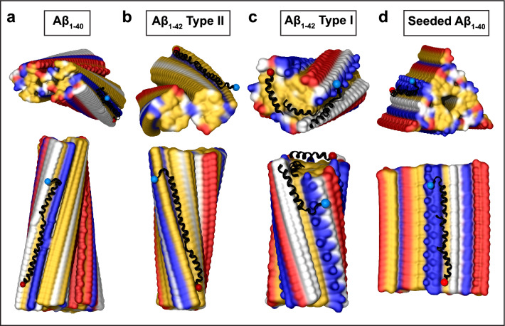
Fig. 5.
Structural models of apoC-III docked onto Aβ fibrils. The models were obtained using apoC-III segments shown in Supplemental Fig. 3b and the fibril structures of Aβ1-40 (a, d) and Aβ1-42 (b, c), PDB ID: 6SHS (a), 7Q4M (b), 7Q4B (c), and 2M4J (d). Blue and red dots indicate N- and C-termini of docked apoC-III. Segment positions are compatible with full-length apoC-III

