FIG. 2.
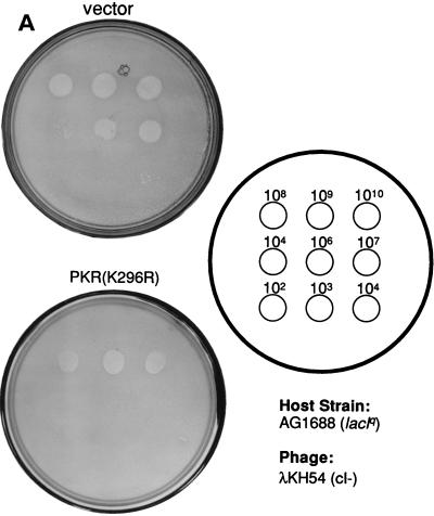
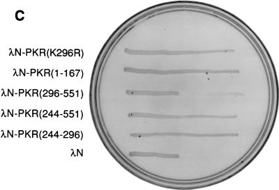
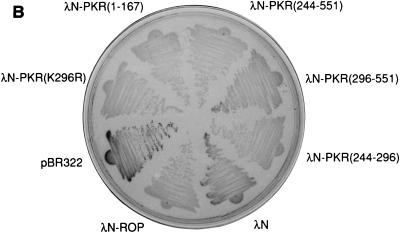
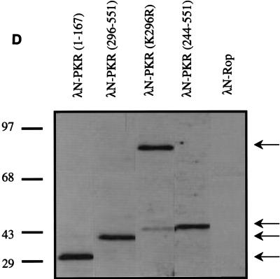
Dimerization properties of λN-PKR fusion proteins. (A) Dot plaque assay. Freshly poured lawns of E. coli (strain AG1688) expressing either λN alone or λN-PKR(K296R) fusion were infected by spotting with 5-μl aliquots of 10-fold dilutions of lysates of λKH54. The titer and pattern of the aliquots are indicated. Sensitivity to λ superinfection was scored by evaluating lysis plaques. (B) Repression of β-Gal expression. Repressor activity of the indicated λN fusions was assessed by expression from the λPR lacZ reporter in strain JH372. A functional λN fusion inhibits lacZ expression, resulting in white colony color. In contrast, cells lacking a functional repressor fusion form blue colonies due to β-Gal production. (C) Cross-streak tests. Phages were striped down the center of an agar plate, and cells harboring the plasmids expressing the indicated λN fusions were streaked perpendicularly across the phage stripe. Sensitive cells were scored by assessing lysis by the phage (i.e., the streak disappeared or was noticeably thinner after crossing the phage zone). (D) Expression of λN chimeric proteins containing different PKR mutants. Crude cell extracts were subjected to SDS-PAGE on a 14% gel and visualized by immunoblot analysis by using a polyclonal antibody raised against PKR (4). Detection of λN-PKR(244–296) was unsuccessful by this analysis. The positions of fusion proteins are indicated by arrows. The molecular size standards are shown in kilodaltons.


