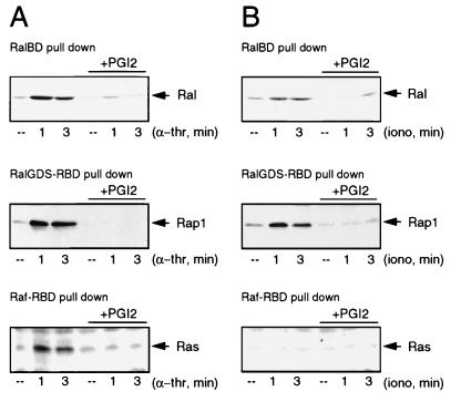
FIG. 6.
Ral activation by Ca2+ correlates with the activation of Rap1 but not Ras. (A) α-Thrombin-induced Ral activation correlates with Rap1 and Ras activation. Platelets preincubated or not for 2 min with 20 ng of PGI2 per ml were stimulated with 0.1 U of α-thrombin (α-thr) per ml for the indicated times. Cell lysates were split and analyzed for the presence of RalGTP, RasGTP, and Rap1GTP. Ral activation (upper panel) was analyzed as described in the legend to Fig. 2. Rap1GTP was isolated with GST-RalGDS-RBD precoupled to glutathione beads and Western blotted with polyclonal anti-Rap1 (middle panel). The lower panel shows Ras activation. RasGTP was isolated with GST-Raf1-RBD, precoupled to glutathione beads, and analyzed by Western blotting with an anti-Ras monoclonal antibody (Transduction Laboratories). These methods for the detection of Ras and Rap1 activation have been described previously (12, 18). (B) Platelets preincubated or not for 2 min with 20 ng of PGI2 per ml were stimulated with 100 nM ionomycin in the presence of 1 mM CaCl2 for the indicated times. Cell lysates were split and analyzed for the presence of RalGTP, Rap1GTP, and RasGTP, as described for panel A. ––, resting platelets.

