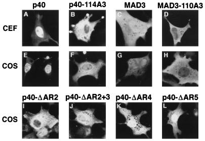
FIG. 2.
Cellular localization of wild-type and mutant IκBα proteins. CEF were transfected with SNV-derived retroviral vectors (A to D) and COS-1 cells were transfected with CMV-derived expression vectors (E to H) encoding either wild-type p40 (A and E), p40-114A3 (B and F), MAD3 (C and G), or MAD3-110A3 (D and H), or COS-1 cells were transfected with SNV-derived expression vectors encoding either p40-ΔAR2 (I), p40-ΔAR2+3 (J), p40-ΔAR4 (K), or p40-ΔAR5 (L). The p40-114A3 protein contains alanine substitutions for leucine 119, leucine 121, and isoleucine 124 in p40. The MAD3-110A3 protein contains alanine substitutions for leucine 115, leucine 117, and isoleucine 120 in MAD3. The p40-ΔAR2 protein contains a deletion of amino acids 98 to 142, encompassing the second ankyrin repeat in p40. The p40-ΔAR2+3 protein contains a deletion of amino acids 117 to 188, encompassing the second and third ankyrin repeats in p40. The p40-ΔAR4 protein contains a deletion of amino acids 189 to 222, encompassing the fourth ankyrin repeat in p40. The p40-ΔAR5 protein contains a deletion of amino acids 223 to 256, encompassing the fifth ankyrin repeat in p40. The cellular localization of the proteins in transfected cells was determined by indirect immunofluorescence with anti-p40 or anti-MAD3 serum. The cells shown are representative of more than 200 cells that were positive for the expression of the indicated proteins (see Table 1 for quantitation).

