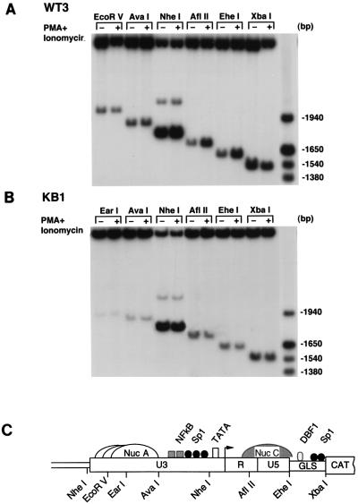
FIG. 3.
NF-κB stimulation induces increased accessibility of the integrated HIV-1 WT-LTR to restriction endonucleases. (A and B) Nuclei isolated from WT3 and KB1 cells maintained in DMEM (−) or stimulated with PMA plus ionomycin (+) were digested with the restriction enzymes indicated at the top. DNA was then purified, digested to completion with BglI and HincII, and analyzed by indirect end labeling. A molecular weight size marker was prepared by digesting a previously described LTR-CAT plasmid, pLCH (15), with several restriction enzymes. (C) Schematic representation of the putative nucleosomal structure and partial restriction map of WT-LTR. Nuc A and Nuc C are positioned relative to micrococcal nuclease and restriction enzyme cleavage sites (11). Nuc A is shown as three adjacent and overlapping structures to indicate that it has not been precisely positioned. Nuc C is represented by a hatched structure to indicate that its accessibility to endonucleases is increased following stimulation with PMA plus ionomycin. GLS, gag leader sequence.

