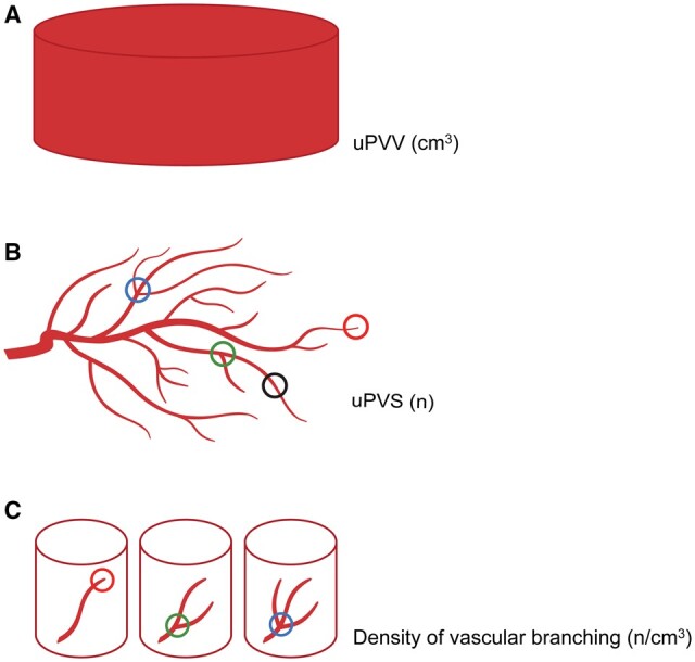
Figure 1.

Graphic representation of virtual imaging markers of first-trimester utero-placental vascular development. (A) The utero-placental vascular volume (uPVV) measures the total volume of all power Doppler signal within the first-trimester placental tissue up to the placental-myometrial interface in cm3. (B) The utero-placental vascular skeleton (uPVS) measures the number and type of branches within the uPVV. Red circle: voxels that are endpoints. Green circle: voxels that are bifurcation points, Blue circle: voxels that are crossing points. Black circle: voxels that are vessel points (no branch or endpoint). In addition, the total vascular length and average vascular thickness are calculated. (C) Density of vascular branching is calculated by dividing the number of endpoints, bifurcation points and crossing points by the uPVV.
