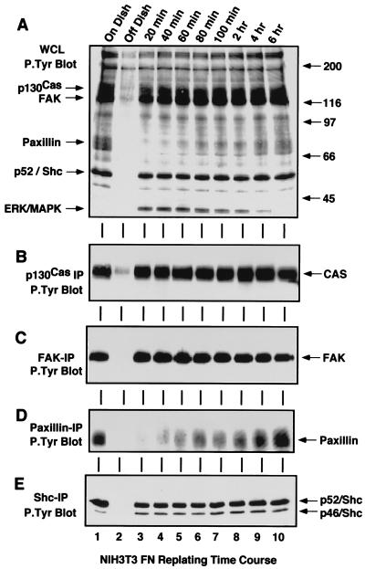
FIG. 1.
Tyrosine phosphorylation of p130Cas, FAK, paxillin, and Shc is not always associated with FN-stimulated signaling events. NIH 3T3 fibroblasts were either serum-starved “On Dish” (lane 1), held in suspension for 30 min “Off Dish” (lane 2), or replated onto FN-coated dishes for the times indicated (lanes 3 to 10). RIPA buffer-treated cell lysates were equalized for protein content and ∼100 μg of WCL (A), or IPs from ∼500 μg of WCL were made with p130Cas (B), FAK (C), paxillin (D), or Shc (E) antibodies. The samples were resolved by SDS-PAGE, analyzed by anti-P.Tyr blotting, and visualized by enhanced chemiluminescence (ECL) detection. Arrows indicate the positions of p130Cas, FAK, paxillin, Shc, and ERK2. ECL exposure time for panel A was 30 s.

