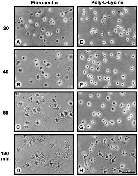
FIG. 3.
Time course of FN-stimulated cell spreading compared to NIH 3T3 cell adhesion to PLL. Serum-starved NIH 3T3 fibroblasts were held in suspension for 30 min and plated (1.3 × 10⁴ cells/cm2) onto either FN-coated (A to D) or PLL-coated (E to H) cell culture dishes. At the times indicated, phase-contrast micrographs were taken with a Nikon inverted microscope (Nikon, Inc., Melville, N.Y.) equipped with a 40× objective lens and photographed with T-MAX 400 film (Kodak). Scale bar, ca. 100 μm.

