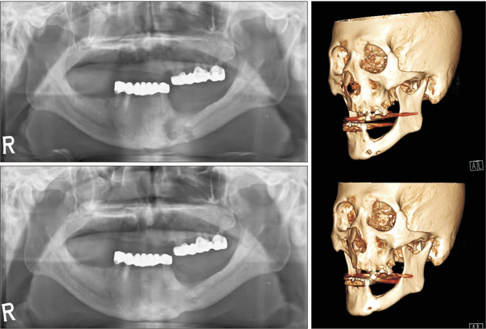
Fig. 2.
Preoperative panoramic view (upper left) shows invasion of the inferior border of the mandible. Postoperative panoramic view (lower left) at seven months shows recovery at the inferior border of the mandible. Reconstructed views of the preoperative (upper right) and postoperative (lower right) lesion.

