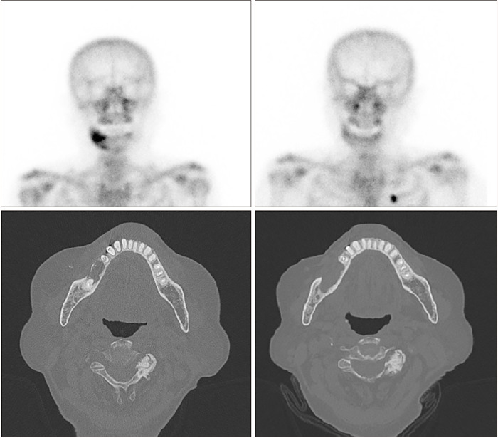
Fig. 5.
Bone scintigraphy at two (upper right) and eight (upper left) months postoperatively. Improved osteonecrosis on the right mandible was confirmed. Computed tomography (CT) showed cortical erosion extending to the mesial surface of the right first premolar area (lower right). Postoperative two-month CT shows normal trabecular bone healing on the debrided area (lower left).

