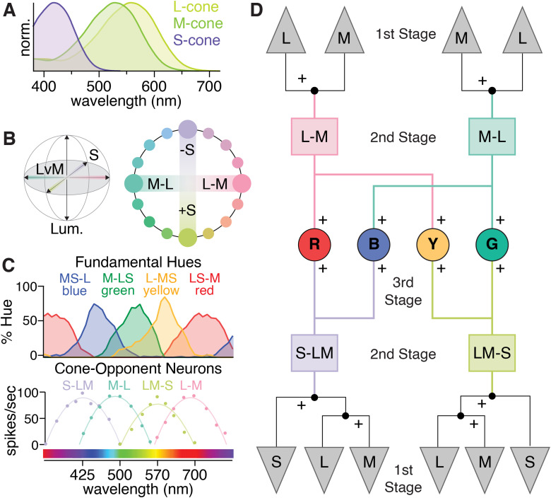
Figure 1.
The most common cone-opponent neurons in the primate early visual system do not match hue perception. A, Normalized spectral sensitivities for L-, M-, and S-cones, which are often called “red,” “green,” and “blue.” However, each is individually colorblind as their outputs confound wavelength with intensity (Baylor et al., 1987). As a result, color vision requires comparing different cone types. B, The isoluminant plane of the three-dimensional DKL color space (gray shading) formed by the cardinal L vs. M and S axes. Blue, yellow, and green are not located along the cardinal axes and thus require both L vs. M and S-cone input. While red is near L–M in the DKL isoluminant plane, S-cone input is needed to explain the redness perceived in short-wavelength violet light. C, Cone-opponent LGN neurons (bottom) initially linked L vs. M to “red–green” and S vs. LM to “blue–green” or “blue–yellow” (De Valois et al., 1966; Wiesel and Hubel, 1966). However, their responses and cone inputs do not align with the tuning curves for red, green, blue, and yellow obtained with hue scaling (top). Adapted from De Valois (2004). D, One form of the multi-stage model, adapted from Stockman and Brainard, 2010. The cardinal directions reflect the second stage consistent with color detection and comparisons of their outputs form a third stage with cone opponency that can explain color appearance. Other multi-stage models omit the once-controversial S-OFF neurons and only use the output of S-ON small bistratified RGCs (De Valois and De Valois, 1993).

