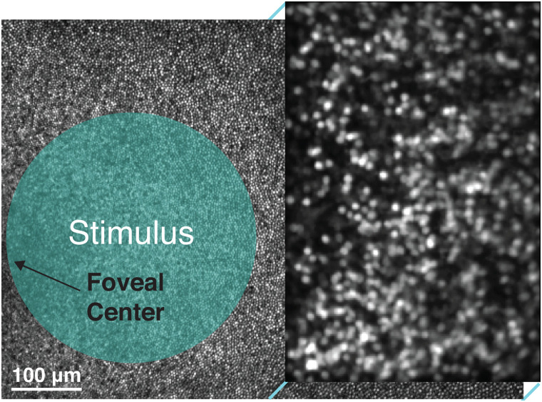
Figure 2.
Experimental paradigm for stimulus delivery and calcium imaging in the living macaque fovea. Diagram illustrating the paradigm for in vivo calcium imaging with AOSLO. Data from M4 is shown with a 3.69 × 2.70° field of view. Reflectance imaging (796 nm) of the cone mosaic was performed across the full field of view while fluorescence imaging (488 nm) was restricted to the right half of the field of view and focused on GCaMP6s-expressing RGCs in the GCL. Visual stimuli were focused on the foveal cones, which are displaced both laterally and axially from the RGCs. Wavefront sensing (847 nm) for real-time detection and correction of the eye's aberrations with adaptive optics is not pictured.

