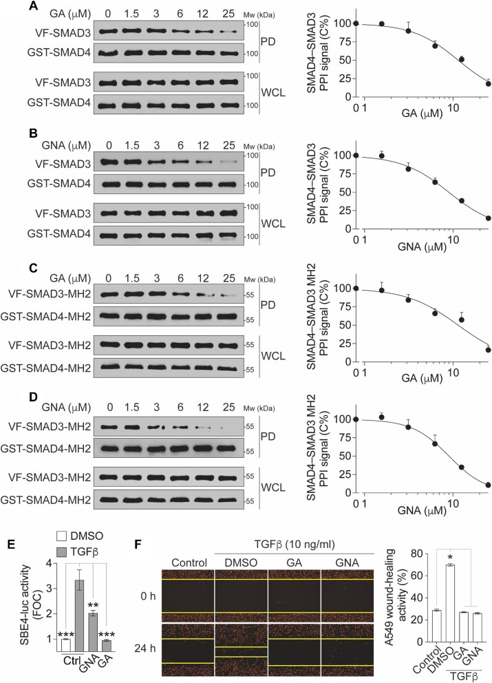
Figure 7.
Confirmation of GA and GNA in GST pull-down and cellular functional assays. (A–D) The inhibition of full-length SMAD4–SMAD3 PPI (A and B) or SMAD4–SMAD3 MH2 domain PPI (C and D) by GA (A and C) or GNA (B and D). The cell lysates expressing GST-SMAD4 and Venus-flag-tagged SMAD3 (VF-SMAD3) (A and B) or GST-SMAD4-MH2 and VF-SMAD3-MH2 (C and D) were treated with compounds as indicated. Left, protein samples from the GST pull-down (PD) and the whole-cell lysates (WCL) were analyzed by western blotting. Right, dose–response curves of the PPI signal were derived from densitometry analysis of the gels. The data are presented as mean ± SEM from three independent experiments. (E) GA and GNA inhibit TGFβ-induced SBE4-luc reporter activity. HEK293T cells expressing endogenous SMAD4 and SMAD3 were treated with TGFβ (10 ng/ml) and/or GA or GNA at 5 μM for 18 h. The TGFβ-induced FOC of the luciferase signals is presented as mean ± SD from three independent experiments. **P < 0.01, ***P < 0.001. (F) GA and GNA inhibit TGFβ-induced migration of A549 cells labeled with NucLight Red fluorescence protein in the nucleus. After wound scratch, cells were treated with TGFβ (10 ng/ml) and/or GA or GNA at 5 μM for 18 h. Left, representative images showing A549 cell (red) migration toward the wound scratch area. Right, the wound-healing activity is quantified and presented as mean ± SD from three independent experiments. *P < 0.05.

