FIG. 6.
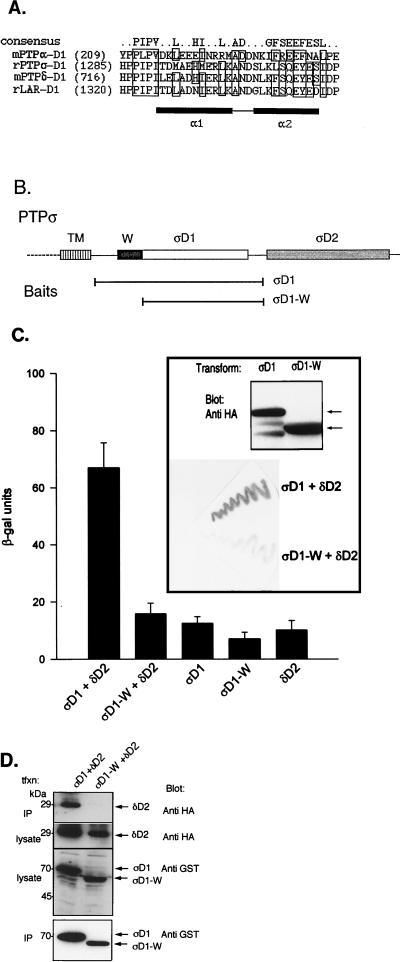
Requirement of the wedge sequence of PTPς-D1 for interaction with PTPδ-D2. (A) Alignment of the wedge (W) sequences of the first catalytic domains of several RPTPs, including mouse (m) RPTPα, rat (r) PTPς, mouse PTPδ, and rat LAR. A more detailed alignment is provided elsewhere (3). Boxed residues represent identical or conserved amino acids conforming to the consensus of the wedge sequence. The α1 and α2 helices are represented by black boxes. (B) Schematic representation of the two-hybrid baits for panel C, including full-length PTPς-D1 (ςD1) or the N-terminally truncated, wedge-deleted (ςD1-W) regions. Both proteins were expressed as fusion proteins with the LexA DNA binding domain. (C) Yeast two-hybrid binding assays of L40 cells cotrans- formed with ςD1 (bait) plus δD2 (prey), with ςD1-W (bait) plus δD2 (prey), or with each construct alone (ςD1, ςD1-W, or δD2). Quantitative β-gal assay results (histograms) are the means ± the standard errors of six determinations. Filter β-gal assay results are shown in the inset (bottom). Levels of protein expression of the PTPς-D1 and PTPςD1-W baits (HA tagged) in these experiments were similar, as determined by immunoblotting with anti-HA antibodies (top of inset, arrows). (D) Lack of coprecipitation of PTPδ-D2 with PTPςD1-W. Cos7 cells were cotransfected with HA-tagged PTPδ-D2 (δD2) together with either GST–PTPς-D1 (ςD1) or the wedge-deleted GST–PTPςD1-W (ςD1-W) construct. Cells were then lysed, and the lysate was incubated with glutathione agarose beads to precipitate GST–PTPς-D1 or GST–PTPςD1-W and associated proteins. Proteins bound to the beads were separated by SDS–10% PAGE and immunoblotted with anti-HA antibodies to test for coprecipitation of PTPδ-D2 (top panel, IP). Aliquots of the lysate of the transfected cells were analyzed for levels of expression of PTPδ-D2 by using anti-HA antibodies (second panel) or GST–PTPς-D1 and GST–PTPςD1-W by using anti-GST antibodies (third panel). The blot in the top panel was then stripped and reprobed with anti-GST antibodies to determine the amount of GST–PTPς-D1 or GST–PTPςD1-W precipitated from the transfected cell lysate (IP, bottom panel). tfxn, transfection.

