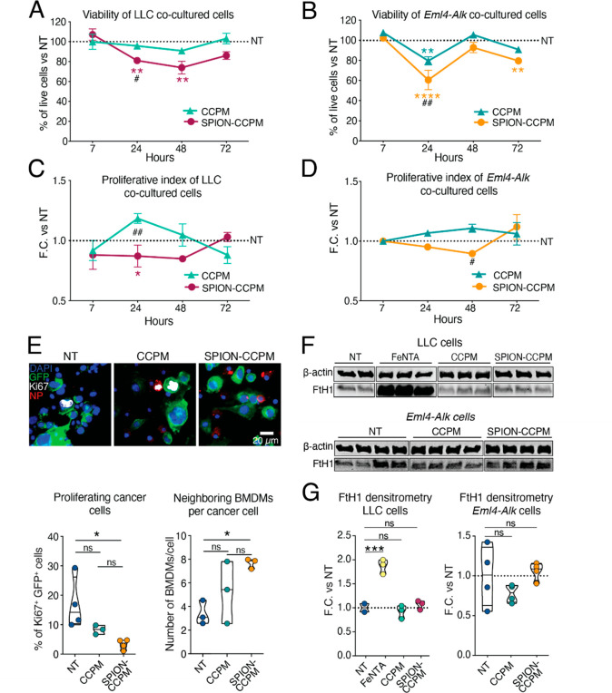
Figure 2.
SPION-CCPM accumulation in macrophages reduces cancer cell proliferation in cocultures. Percentage of viable Lewis lung carcinoma (LLC) cells (A) and Eml4-Alk cells (B) cocultured with bone marrow-derived macrophages (BMDMs) after treatment with or without CCPMs or SPION-CCPMs. Proliferative index of LLC cells (C) and Eml4-Alk cells (D) maintained in coculture with BMDMs represented as a fold change (F.C.) vs nontreated (NT) cells. (E) Ki67 immunofluorescence in cocultured cells treated with or without CCPMs or SPION-CCPMs (NP) after 24 h. Cancer cells are endogenously labeled with GFP. Quantification of percentage of Ki67+ cancer cells in 3000 cells obtained from three mice (below, left) and the number of neighboring BMDMs per cancer cell (below, right). (F) Western blot of the FtH1 protein in cancer cells cocultured with BMDMs treated with or without FeNTA, CCPMs, or SPION-CCPMs after 48 h. (G) Densitometry analyses of the Western blots in (F). #SPION-CCPM vs CCPM comparison; *significance of indicated colored condition vs NT condition; *p < 0.05, **p < 0.005, ***p < 0.0005, ****p < 0.0001; Two-way ANOVA (A, B, C, D, G) or one-way ANOVA (E, G). For all, at least n = 3.

