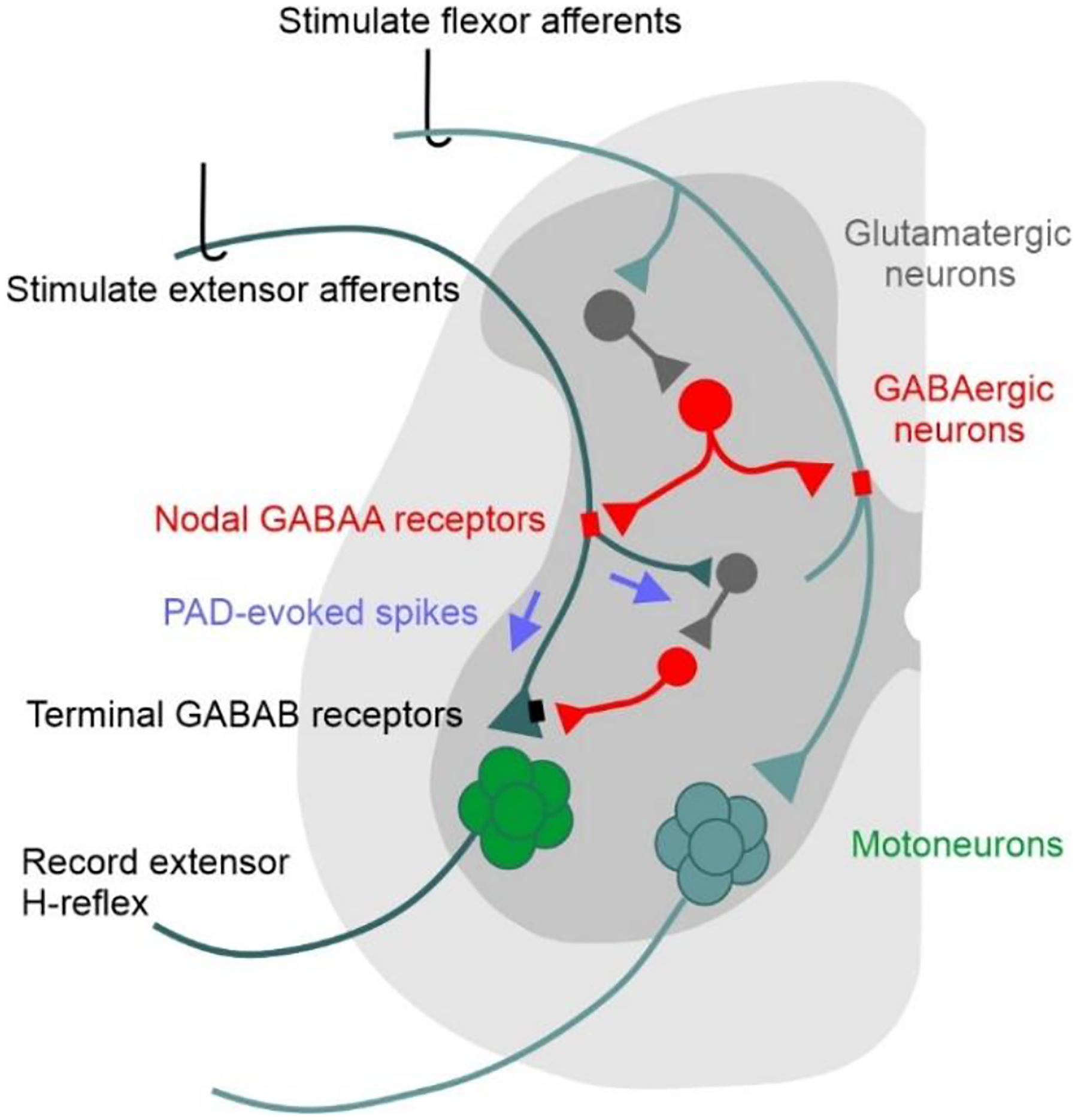
Figure 11. Schematic of PAD pathways mediating post-activation depression in the spinal cord.

Activation of flexor afferents (cyan) activates a tri-synaptic dorsal PAD pathway (glutamatergic interneuron dark grey, GABAaxo neuron red) with axo-axonic projections to an extensor afferent (dark green), activating nodal GABAA receptors (red) and PAD in the extensor afferent. If the resulting PAD in the extensor afferent reaches sodium spiking threshold, PAD-evoked spikes (purple arrows) travel orthodromically to: 1) depolarize extensor afferent terminals, evoking an EPSP in the extensor motoneurons and a subsequent MSR/H-reflex; 2) activate a trisynaptic circuit containing GABAergic neurons that activate GABAB receptors (black) on the extensor afferent terminal. Thus, the PAD-evoked spike in the extensor Ia afferent can produce early and late post-activation depression of subsequently activated MSRs/H-reflexes by: 1) decreased afferent excitability from collision of orthodromic action potentials with antidromic PAD-evoked spikes; 2) transmitter depletion following the PAD-evoked spike entering the extensor Ia afferent; and/or 3) inhibition of the afferent terminals by GABAB receptors indirectly activated by the PAD-evoked spike (GABAB mediated presynaptic inhibition).
