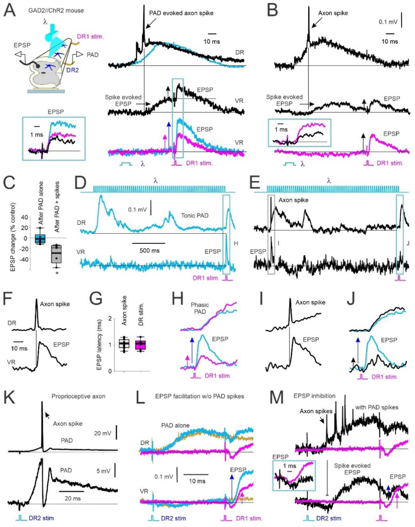
Figure 3: GABAA mediated PAD in Ia afferents with and without spiking and subsequent EPSPs.

A) Left, In vitro experimental set up showing PAD recorded from dorsal root 2 (DR2) activated by light (λ) in a GAD2//ChR2 positive mouse. Monosynaptic reflex (MSR) activated by stimulation of DR1, subsequent EPSPs recorded from ventral root (VR). Right, top: PAD recorded from DR2 with (black) and without (blue) PAD-evoked axon spike. Middle: VR recording of evoked EPSP from the PAD-evoked axon spike in top trace. Subsequent test EPSP activation (DR1 stimulation) is reduced (vertical arrows indicate EPSP amplitude). Bottom: EPSP from DR1 stimulation alone (pink) compared to EPSP following the light-evoked, non-spiking PAD shown in top trace (blue). Box: Overlay of EPSPs from middle and bottom traces in A. B) Same as in A but test EPSP from DR1 stimulation delivered at a longer interval after the PAD spike-evoked EPSP returned to baseline. Box: Overlay of conditioned (black) and control (pink, DR1 stimulation only) EPSPs. C) Change in amplitude of light-conditioned EPSPs (100 – 150 ms ISI) after a non-spiking PAD (PAD alone) and after a PAD with spike(s) as a percentage of an EPSP evoked from DR1 stimulation alone (control). Box plots: median, thin line; mean, thick line; interquartile range IQR, box bounds; most extreme data points within 1.5 × IQR, standard error bars. EPSPs after a PAD-evoked spike were smaller than a 0% change (* p = 0.021) but not after a non-spiking PAD ended (After PAD alone) (p = 0.151, Mann-Whitney U test, n=5 mice). D-E) Top: Light-evoked tonic PAD recorded from DR2 (middle trace) without (D, blue) and with (E, black) a PAD-evoked spike. Bottom: VR recordings of associated of monosynaptic EPSP from DR1 stimulation and PAD spike-evoked EPSP. F) Example DR and VR recording of a PAD-evoked axon spike and resulting EPSP at a monosynaptic latency on expanded time scale. G) Comparison of EPSP latency following PAD evoked axon spike and DR 1 stimulation (not significantly different, p = 0.683, Student’s t test, n = 6 mice). H-J) Expanded view of PAD (top) and EPSPs (bottom) evoked in D and E. Similar results observed in in n=6/6 mice. K-M) Same as in A and B but DR2 stimulation (1.5 × T) used to evoke PAD instead of light. Similar results in n = 5/5 rats. K) Top: Rat intracellular (IC) recording of proprioceptive (Ia) afferent with PAD-evoked axon spike from DR2 stimulation. Bottom: expanded vertical axis of top trace. L-M) PAD evoked with DR2 stimulation. Top: DR recording. Bottom: VR recording. L) PAD evoked without axon spikes. Pink: EPSP alone from DR1 stim. Blue: Non-spiking PAD evoked from DR2 stimulation and facilitated EPSP from DR1 stimulation. Yellow: PAD from DR2 stimulation applied alone. M) PAD, axon spikes and motoneuron EPSPs evoked from DR2 stimulation and test EPSP from DR1 stimulation without (pink) and with (black) spiking PAD (EPSPs expanded in box).
