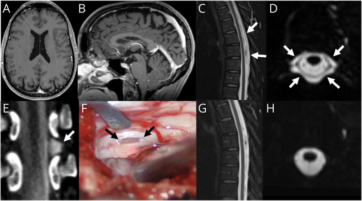
Figure 2. “Normal” Brain MRI in a Patient With SIH.
(A) Axial T1 postcontrast MRI demonstrates no pachymeningeal enhancement or subdural collections. (B) Sagittal T1 MRI demonstrates no significant sagging of the brainstem. (C) Sagittal T2 STIR MRI demonstrates a posterior epidural fluid collection. (D) Axial 3D T2 fat-saturated image demonstrates abnormal epidural fluid surrounding the dura in the midthoracic spine. (E) Dynamic decubitus myelography demonstrates extravasation of subarachnoid contrast into the lateral epidural space at T9-T10 (arrow). (F) Intraoperative photograph during surgical repair demonstrates a lateral dural defect at T9-T10 along the axilla of the left T9 nerve root (arrows). Postoperative T2 fat-saturated imaging in the sagittal (G) and axial (H) planes shows resolution of the epidural fluid collection.

