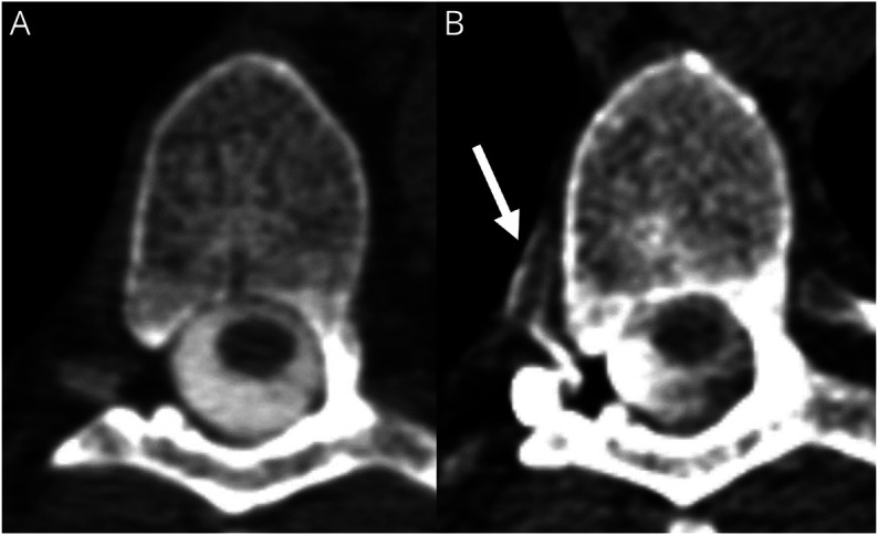
Figure 3. Conventional vs Decubitus Myelography in a Patient With CSF Venous Fistula.

(A) Conventional myelogram at T6-7 demonstrates no evidence of CSF leak or CVF. (B) Dynamic decubitus CT myelography performed in the same patient demonstrates a CSF-venous fistula at T6-7 (arrow).
