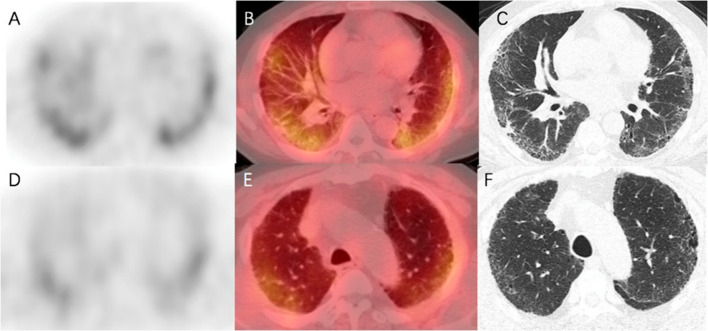
Fig. 7.
Fused axial SPECT/CT images in a patient affected by idiopathic pulmonary fibrosis. Upper row: corresponding emissive A, fused B and transmissive C slices of the lower left and right pulmonary lobes; note the intense tracer incorporation. Lower row: lower tracer incorporation can be observed in the corresponding emissive D, fused E and transmissive F slices of the middle-upper region of the lungs. Reprinted from (Liu et al. 2023b), under a Creative Commons Attribution 4.0 International License (http://creativecommons.org/licenses/by/4.0/). No changes were made

