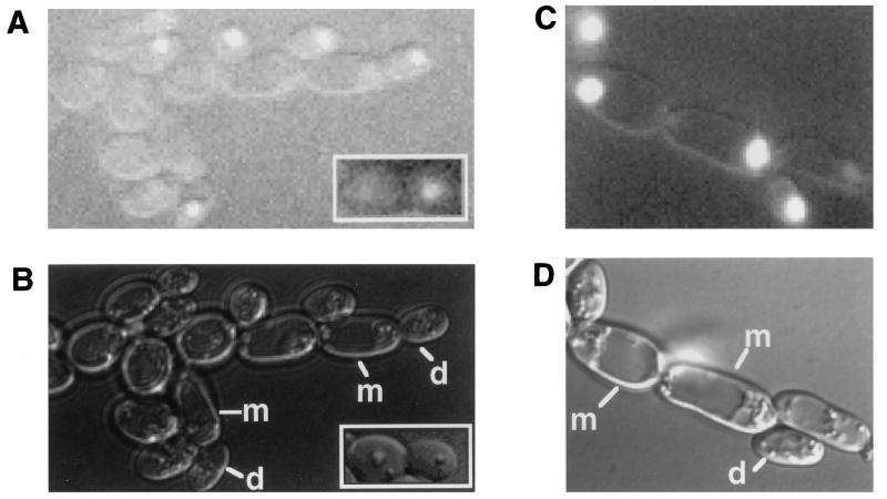
FIG. 6.
Localization of GFP-Ash1 during pseudohyphal growth. Fluorescent (A and C) and differential interference contrast (B and D) views of cells are shown. (A and B) Diploid strain SC126, which is heterozygous for GFP-ASH1, grown on SLAD for 12 h. Insets show a mother-daughter pair of yeast form cells of the same diploid strain grown on SD-Ura. (C and D) Diploid ash1Δ/ash1Δ strain SC125 with pNC513, which expresses GFP-ASH1 from the 2μm vector pRS426, grown on SLAD for 12 h. m, mother cell; d, daughter cell.

